
RHEUMATOID ARTHRITIS
| A | Juxta-articular osteosclerosis | |
| B |
Bamboo spine |
|
| C |
Bone erosions |
|
| D |
Peri-articular calcification |
Which one of the following is a recognized X-Ray feature of rheumatoid arthritis?
| A |
Juxta-articular osteosclerosis |
|
| B |
Bamboo spine |
|
| C |
Bone erosions |
|
| D |
Peri-articular calcification |
Rheumatoid arthritis is an autoimmune disease characterized by symmetric progressive polyarthritis.
Inflammation of synovium cause cartilage destruction, and bone erosion. Bone erosion is the characteristic x-ray finding in a case of rheumatoid arthritis(RA).
Ref: Physical Medicine and Rehabilitation Board Review By Sara J. Cuccurullo, Page 111; Examination Medicine By Nicholas Tailey, Simon O’Connor, Page 347; Differential Diagnosis in Conventional Radiology By Francis A. Burgener, 3rd Edition, Page 192-4.
| A | Both Assertion and Reason are true, and Reason is the correct explanation for Assertion | |
| B |
Both Assertion and Reason are true, and Reason is not the correct explanation for Assertion |
|
| C |
Assertion is true, but Reason is false |
|
| D |
Assertion is false, but Reason is true |
Ans.C
“Swan neck” deformity in rheumatoid arthritis is due to the flexion of the DIP joint with an extension of the PIP joint.
Other deformities are an ulnar deviation of the fingers (the most common), boutonniere deformity (hyperextension of the DIP joint with flexion of the PIP joint), valgus deformity of the knee, and volar subluxation of the MTP joints.
The primary goal of glucocorticoid treatment in rheumatoid arthritis is –
| A |
Suppression of inflammation and improvement in functional capacity |
|
| B |
Reversal of the degenerative process |
|
| C |
Development of a sense of well-being in the patient |
|
| D |
Prevention of suppression of the hypothalamic pituitary-adrenal axis |
Ans. is ‘a’ i.e., Suppression of inflammation and improvement in functional capacity
All of the following are features of systemic Juvenile Rheumatoid Arthritis except-
| A |
Uveitis |
|
| B |
Rash |
|
| C |
Fever |
|
| D |
Hepatosplenomegaly |
Ans.(A) Uveitis
The eye manifestations are seen in Pauciarticular and Polyarticular JRA but not in systemic JRA.
Which one of the following is a recognized X-Ray feature of rheumatoid arthritis?
| A |
Juxta-articular osteosclerosis |
|
| B |
Sacroilitis |
|
| C |
Bone erosions |
|
| D |
Peri-articular calcification |
C i.e. Bone Erosion
All inflamatory arthritis share a lot of radiological features in common. Rheumatoid arthritis is differentiated from other inflammatory seronegative arthropathies by symmetricalQ, small joint (esp MCP & PIP joints) involvementQ, more osteoporosis & bony erosionsQ. In seronegative arthritis, there is asymmetrical, large joint (esp SI, spine & DIP joint of hand) involvement, more periostitis & less osteoporosis.
All the following are true about Rheumatoid arthritis except:
| A |
Positive for Anti-IgG antibody |
|
| B |
Juxta-articular osteoporosis |
|
| C |
Morning stiffness |
|
| D |
C Reactive protein indicates better prognosis |
Answer is D (C Reactive protein indicates better prognosis)
Persons who present with high titers of Rheumatoid factor, C Reactive protein and Haptoglobin have a worse prognosis than the individuals who present with Subcutaneous nodules or Radiographic evidence of erosions
All of the following are true about Rheumatoid Arthritis, Except
| A |
PIP and DIP Joints involved equally |
|
| B |
Pathology limited to articular cartilage |
|
| C |
20 percent of patients have extra articular manifestations |
|
| D |
All |
Answer is A, B, C(PIP and DIP Joints involved Tally; Pathology Limited to articular cartilage; 20 percent have extra articular manifestations)
- Women are affected 3 times more commonly than men
- Pathology is not limited to articular cartilage alone
- The majority of destruction occurs in juxtaposition to the inflamed synovium, or pannus, that spreads to cover the articular cartilage’
- The precise mechanism by which bone and cartilage destruction occurs has not been completely resolved.
- Although the synovial fluid contains a number of enzymes potentially able to degrade cartilage, the majority of destruction occurs in juxtaposition to the inflamed synovium, or pannus, that spreads to cover the articular cartilage.
- The potential of the synovial inflammation to cause cartilage damage and bone erosions and subsequent changes in joint integtrity is the hallmark of the disease
- PIP and DIP Joints are not involved equally (DIP jolts are usually/typically spared)
- ‘RA most often causes symmetric arthritis with characteristic involvement of certain specific joints such as the proximal interphalangeal and metacarpophalangeal joints.
- The distal interphalangeal joints are rarely involved’.
- Extra articular manifestations are seen in upto 40 percent of patients
- ‘It is estimated that us many as 40% ofputients may have extraarticular manifestations, and in -15% these are severe’
- Rheumatoid nodules are seen in approximately 20% of patents
- Rheumotoid nodulels may develop in 20-30% of persons with RA’
Which part of the spine is most commonly affected in Rheumatoid arthritis:
| A | Cervical | |
| B |
Lumbar |
|
| C |
Thoracic |
|
| D |
Sacral |
Answer is A (Cervical):
Rheumatoid arthritis commonly involves the joints of hands, wrist, elbow, knees, ankle, and feet in a symmetrical manner.
Axial skeleton involvement is usually limited to Upper Cervical Spine.
Which radiological feature would help differentiate rheumatoid arthritis with SLE?
| A |
Erosion |
|
| B |
Juxta-articular osteoporosis |
|
| C |
Subluxation of MCP joint |
|
| D |
Swelling of PIP joint |
Answer is A (Erosion):
SLE is characterized by non erosive arthritis while bone erosions are characteristic in Rheumatoid Arthritis.
Rheumatoid factor in Rheumatoid arthritis is:
| A |
IgG |
|
| B |
IgM |
|
| C |
IgA |
|
| D |
IgD |
Answer is B (IgM):
Rheumatoid arthritis is an IgM antibody directed against the Fe fragment of IgG.
Rheumatoid factor in rheumatoid arthritis is important because:
| A |
RA factor is associated with bad prognosis |
|
| B |
Absent RA factor rules out the diagnosis of Rheumatoid arthritis |
|
| C |
It is very common in childhood Rheumatoid arthritis |
|
| D |
It correlates with disease activity |
Answer is A (R. A. factor is associated with bad prognosis) :
Rheumatoid factor is only of prognostic significance because patients with high titres tend to have more severe
and progressive disease with extraarticular manifestations
- Rheumatoid factors are autoantibodies of IgMQ type that react with FC portion of IgGQ
- The presence of RA factor is not specific for R.A.
- The presence of Rheumatoid factor does not establish the diagnosis of RA
| A | Anti-CCP Antibodies | |
| B |
IgA Rheumatoid Factor |
|
| C |
IgG Rheumatoid Factor |
|
| D |
IgM Rheumatoid Factor |
Answer is A (Anti-CCP Antibodies):
‘Anti CCP antibodies are the most specific blood test for rheumatoid arthritis (specificity –95%)’ – CMDT 09/727
Anti-cyclic citrullinated peptide (anti-CCP) antibody testing is particularly useful in the diagnosis of rheumatoid arthritis, with high specificity,
presence early in the disease process, and ability to identify patients who are likely to have severe disease and irreversible damage.
Anti-CCP antibodies have not been found at a significant frequency in other diseases to date, and are more specific than rheumatoid factor for detecting rheumatoid arthritis.
Type of anemia seen in Rheumatoid arthritis is:
| A |
Microcytic hypochromic anaemia |
|
| B |
Macrocytic hypochromic anaemia |
|
| C |
Normocytic hypochromic anaemia |
|
| D |
Normocytic normochromic anaemia |
Answer is D (Normocytic normochromic anaemia):
Normocytic normochromic anaemia is frequently present in Rheumatoid arthritis. It is thought to reflect ineffective erythropoesis, large stores of iron are found in the bone marrow’–
Causative agent for rheumatoid arthritis is:
| A |
Mycoplasma |
|
| B |
Mycobacaterium avium |
|
| C |
Yersinia |
|
| D |
Herpes virus |
Answer is A (Mycoplasma)
The true causative agent for Rheumatoid arthritis is not known. Mycoplasma has been suggested as a possible causative agent, and hence is the single best answer of choice
All of the following are true about Rheumatoid Arthritis, Except:
| A |
HLA-DR determine genetic susceptibility |
|
| B |
HLA-B27 determine genetic susceptibility |
|
| C |
Anemia |
|
| D |
Subcutaneous nodules |
Answer is B (HLA-B27 determine genetic susceptibility):
HLA-B27 antigen is associated with seronegative spondyloarthropathies and not Rheumatoid Arthritis. RA is associated with HLA DR alleles and may present with anemia, subcutaneous nodules and joint deformities.
HLA B 27 associated seronegative spondyloarthropathies
- Ankylosing spondylitis
- Psoriatic arthritis
- Reactive arthritis (Reiter’s syndrome)
- Enteropathic arthritis (Associated with Inflammatory Bowel disease)
- Undifferentiated spondyloarthropathy
September 2012
| A | Carpal tunnel syndrome | |
| B |
Presence of deformities |
|
| C |
Mononeuritis multiplex |
|
| D |
Involvement of articular cartilage |
Ans. C i.e. Mononeuritis multiplex
Felty’s syndrome is associated with which of the following?
| A |
Osteoarthritis |
|
| B |
Rheumatoid arthritis |
|
| C |
Ankylosing spondylitis |
|
| D |
Psoriatic arthritis |
Ans. B: Rheumatoid Arthritis
Felty syndrome
- Felty syndrome is a rare hematological syndrome in rheumatoid arthritis.
Epidemiology
- It is thought to occur in ~ 1% of patients with rheumatoid arthritis.
Clinical presentation
- Felty syndrome comprises of the triad of:
1. Rheumatoid arthritis
- typically with severe articular and extra-articular disease
2. Splenomegaly
3. Neutropenia
Pathology
Rheumatoid factor (RF): >95% of patients are positive .
Antinuclear antibody (ANA): 47-100% are positive.
HLA-DR4*0401 antigen: 78% of patients have the antigen.
Large granular lymphocyte (LGL) expansion: ~ 30% of patients may have it .
Treatment and prognosis
- Treatment is through immunosuppression to treat Felty syndrome and rheumatoid arthritis, such as use of Methotrexate and Rituximab.
- If frequent severe bacterial infections, G-CSF may be administered.
September 2011
| A | Symmetric swelling | |
| B |
Morning stiffness |
|
| C |
Positive Rheumatoid factor |
|
| D |
Swelling of any 2 joints of the bodies |
Ans. D: Swelling of any 2 joints of the bodies
New diagnostic criteria for rheumatoid arthritis (1987) includes 1) morning stiffness, 2) swelling of 3 or more specified joints, 3) swelling of joint(s) in the hands and wrist, 4) symmetrical swelling, 5) rheumatoid nodule, 6) rheumatoid factor positive and 7) X-ray changes – erosion or unequivocal peri-articular osteopenia
If four or more of these are present, it is rheumatoid arthritis
| A |
Decreased joint space |
|
| B |
Juxta articular osteoporosis |
|
| C |
Periarticular soft tissue swelling |
|
| D |
Suhchondral erosions |
Ans. Periarticular soft tissue swelling
Swan neck deformity is seen in ‑
| A |
Osteoarthritis |
|
| B |
Rheumatoid arthritis |
|
| C |
Psoriatic arthritis |
|
| D |
Gout |
Ans. is b’ i.e., Rheumatoid arthritis
Important deformities of hand in RA
- Boutonniere deformity : Flexion contracture of PIP joint and extension of DIP joint.
- Swan neck deformit: Hyperextension of PIP joint and flexion at DIP joint.
- Z-deformity : Radial deviation of wrist with ulnar deviation of fingers.
- Hyperextension of 1st interphalangeal joint and flexion of MP joint.
| A | Heberden nodes | |
| B |
Swan neck deformity |
|
| C |
Ulnar deviation of fingers at metacarpopalyngeal joint |
|
| D |
Symmetric reduction of joint space |
Ans. is ‘a’ i.e., Heberden nodes
Heberden nodes is a feature of osteoarthritis and not rheumatoid arthritis.
Important hand deformities of hand in RA
- Boutonniere deformity : Flexion contracture of PIP joint and extension of DIP joint.
- Swan neck deformity : Hyperextension of PIP joint and flexion at DIP joint.
- Z-deformity : Radial deviation of wrist with ulnar deviation of fingers.
- Hyperextension of 1st interphalangeal joint and flexion of MP joint.
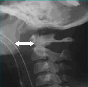
| A | Rheumatoid arthritis. | |
| B |
Osteoarthritis. |
|
| C |
Fibrous dysplasia. |
|
| D |
Ankylosing spondylitis. |
The deformity as shown in the picture above represents Atlanto-axial subluxation, which is most characteristic of the Rheumatoid arthritis.
Rheumatoid arthritis (RA) is an autoimmune disease that causes chronic inflammation of the joints. Autoimmune diseases are illnesses that occur when the body’s tissues are mistakenly attacked by their own immune system. The immune system contains a complex organization of cells and antibodies designed normally to “seek and destroy” invaders of the body, particularly infections. Patients with autoimmune diseases have antibodies and immune cells in their blood that target their own body tissues, where they can be associated with inflammation. While inflammation of the tissue around the joints and inflammatory arthritis are characteristic features of rheumatoid arthritis, the disease can also cause inflammation and injury in other organs in the body. Because it can affect multiple other organs of the body, rheumatoid arthritis is referred to as a systemic illness and is sometimes called rheumatoid disease. Rheumatoid arthritis is a classic rheumatic disease.
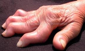
Deformity as shown in the picture below is characteristic of which among the following ?
| A |
Osteoarthritis. |
|
| B |
Rheumatoid arthritis. |
|
| C |
Ankylosing spondylitis. |
|
| D |
Psoriatic arthritis. |
Deformity as shown in the picture above represents Swan neck deformity, which is characteristic of Rheumatoid arthritis.
Swan neck deformity is a deformed position of the finger, in which the joint closest to the fingertip is permanently bent toward the palm while the nearest joint to the palm is bent away from it (DIP flexion with PIP hyperextension).
All of the following are true about Rheumatoid arthritis, except
| A |
PIP and DIP joints involved equally |
|
| B |
Pathology limited to articular cartilage |
|
| C |
Women are affected 3 times more commonly than men |
|
| D |
20% of patients have extra articular manifestations |
Ans. is ‘d’ i.e., 20% of patients have extra articular manifestations
Association of Rheumatoid Arthritis with HLA DR-4
- The class II major histocompatibility complex allele HLA-DR4 and related alleles are known to be a major genetic risk factor for Rheumatoid arthritis.
- Rheumatoid Arthritis is strongly associated with HLA DR4
- The genetic risk for Rheumatoid arthritis is associated with allelic variation in the HLA-DRBI “gene i.e. DRB1 0401, 04, 05”.
- Some of the HLA DRB1 alleles bestow a high risk of disease
Clinical features:
- It occurs between the age of 20 to 50 years.
- Women are affected about 3 times more commonly than men.
- Following presentations are common:
a) An acute, symmetrical polyarthritis:
- Pain and stiffness in multiple joints (at least four)
- Symptoms of articular inflammation.
PIP joints of fingers
Wrists, knees, elbows, ankles

| A | Psoriatic Arthritis | |
| B |
Rheumatoid Arthritis |
|
| C |
Osteoarthritis |
|
| D |
Relapsing polychondritis |
Ans:B.)Rheumatoid Arthritis.
Image shows: Severe rheumatoid arthritis of the wrist and hand. The ulnar styloid is destroyed with the rest of the distal ulna (white arrow), there is destruction of the carpal bones (yellow arrow) and there is dislocation of the 1st metacarpal on the destroyed trapezium (blue arrow). All of the bones are osteopenic.
RHEUMATOID ARTHRITIS
- Most common inflammatory arthritis in women
- Symmetric peripheral polyarthritis
- Female gender, cigarette smoking are risk factors
- Strongly associated with HLA-DR4
Pathogenesis:
- Accumulation of neutrophils in the synovial fluid and along the synovial surface(not in the synovial stroma)
- Aggregation of organizing fibrin covering the portions of synovium and floating in the joint space as rice bodies
- Pannus formation – a mass of synovium and synovial stroma consisting of inflammatory cells, granulation tissue and fibroblasts which causes erosion and destruction of articular cartilage (principle target) eventually resulting in ankylosis
Clinical features:
- Early morning joint stiffness lasting more than 1 hour and easing with physical activity
- Earliest involved joints are typically the small joints of the hands and feet
- Wrists, metacarpophalangeal (MCP), proximal interphalangeal (PIP) joints are most frequently involved
- Subcutaneous rheumatoid nodules, most commonly situated over bony prominences but also observed in the bursae and tendon sheaths
Joints involved:
- Symmetrical involvement of MCP, PIP and MTP
- Large joint involvement is common but occurs later
- Spine is spared except the cervical spine which is commonly involved
Deformities:
- Flexor tendon tenosynovitis is a frequent hallmark of RA and leads to decreased range of motion, reduced grip strength, and trigger fingers
- Swan neck deformity– hyperextension of PIP with flexion of DIP
- Button hole or boutonniere deformity – flexion of the PIP joint with hyperextension of the DIP
- Z-line deformity of the thumb – subluxation of the first MCP with hyperextension of the first interphalangeal (IP) joint
- Ulnar deviation of fingers – subluxation of MCP, with subluxation of the proximal phalanx to the volar side of the hand
- Cock-up toe – dorsal subluxation of MTP
- Piano-key movement of the ulnar styloid – inflammation about the ulnar styloid and tenosynovitis of the extensor carpi ulnaris
- Pes planovalgus (flat feet)
- Hammer toe & Wind – sweep deformities of toes
- Late triple knee deformity
Laboratory features:
- Synovial fluid WBC counts: 5000 – 50,000 WBC/μ3(
Radiological features
- Juxtaarticular osteopenia
- Bone erosions (often first evident at the ulnar styloid and at the juxtaarticular margin)
- Narrowing of joint space due to loss of articular cartilage
Rheumatoid factor
- Autoantibody, usually IgM
- Not specific for RA (absence cannot exclude RA); Positive in 70-90% patients
- Positive RF is associated with worse prognosis
- Level parallels the activity of the disease
- Anti-CCP antibody more specific than Rheumatoid factor in the diagnosis of RA
- Anti-CCP positivity – worse prognosis
Criteria for diagnosis of RA (four or more of the following)
- Morning stiffness > 1hr
- Arthritis of 3 or more joints
- Arthritis of hand joints
- Rheumatoid nodules
- Rheumatoid factor
- Radiological changes
- Duration of 6 weeks or more
Extra articular manifestations – more common under the following conditions
- History of smoking
- Early onset of significant physical disability
- Test positive for serum RF
- Ocular
- Scleritis, episcleritis
- Scleromalacia
- Keratoconjunctivitis sicca
- Felty’s syndrome
- Nodular RA
- Splenomegaly
- Neutropenia
- Amyloidosis
- Cardiac
- Pericarditis(MC),myocarditis,endocarditis
- Pericardial effusion
- Conduction defects
- Vasculitis
- Pyoderma gangrenosum
- Mononeuritis multiplex
- Pulmonary
- Nodules (MC)
- Fibrosing alveolitis
- Bronchilitis
- Exudative Pleural effusion with low glucose and pH
- Caplan’s syndrome
- Coal worker’s pneumoconiosis
- RA
Treatment:
- DMARDs- methotrexate (first choice) hydroxychloroquine, sulfasalazine, leflunomide
- NSAIDs, Corticosteroids
- Anti-TNF-α: Etarnacept, Infliximab, Certolizumab
- Anakinra (anti-IL-1), abatacept, monoclonal antibody(rituximab, tocilizumab)
