STAGES OF ANESTHESIA
Stages of anaesthesia were described by Guedel with
| A | Ether | |
| B | Chloroform | |
| C |
N20 |
|
| D |
Halothane |
Stages of anaesthesia were described by Guedel with
| A |
Ether |
|
| B |
Chloroform |
|
| C |
N20 |
|
| D |
Halothane |
Ether
Which of the following technique is used to evaluate the intraoperative awareness of an individual during anaesthesia?
| A |
Cerebral pulse oximetry |
|
| B |
End tidal C02 |
|
| C |
Bispectral index |
|
| D |
Colour Doppler |
- BIS number < 60: Patient unable to respond to verbal commands
- BIS number > 70: Corresponds to a higher likelihood of awareness
- BIS number of 90- 100: Awake
BIS numbers between 45 and 60 are, in general, considered to be optimal for a relatively healthy patient undergoing a routine general anesthetic and surgery.
Ref:Clinical Anesthesiology By G. Edward Morgan, 4th Edition, Chapter 6
Stages of anesthesia were established by
| A |
Ether |
|
| B |
N20 |
|
| C |
Halothane |
|
| D |
Chloroform |
A i.e. Ether
From the following given options, contributions from this famous Anesthesiologist as shown in the image is?

| A |
Stages of Anesthesia |
|
| B |
Oropharyngeal Airway |
|
| C |
Discovering Oxygen |
|
| D |
Both a and b |
Ans:D-Both a and b. Stages of Anesthesia and Oropharyngeal Airway.
The image shown is of Anesthesiologist: Arther Guedel
Arther Guedel
- Arthur Ernest Guedel (June 13, 1883 – June 10, 1956) was an American anesthesiologist.
- He designed Stages of Anesthesia and oropharyngeal Airway.
- Guedel’s classification is a means of assessing of depth of general anesthesia
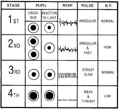
Stages of Anaesthesia
- Stage I (Stage of Analgesia or the stage of Disorientation): from the beginning of induction of anesthesia to loss of consciousness.
- Stage II (Stage of Excitement or the stage of Delirium): from loss of consciousness to onset of automatic breathing. Eyelash reflex disappears but other reflexes remain intact and coughing, vomiting and struggling may occur; respiration can be irregular with breath-holding.
- Stage III (Stage of Surgical anesthesia): from the onset of automatic respiration to respiratory paralysis. It is divided into four planes:
- “Plane I” – from the onset of automatic respiration to the cessation of eyeball movements.
- “Plane II” – from the cessation of eyeball movements to the beginning of paralysis of intercostal muscles.
- “Plane III” (Surgical Anaesthesia) from beginning to completion of intercostal muscle paralysis.
- “Plane IV” – from complete intercostal paralysis to diaphragmatic paralysis (apnoea).
- Stage IV: from the stoppage of respiration till death. Anaesthetic overdose causes medullary paralysis with respiratory arrest and vasomotor collapse. Pupils are widely dilated and muscles are relaxed.
Oropharyngeal airway (also known as an oral airway, OPA or Guedel pattern airway)
- It is a medical device called an airway adjunct used to maintain or open a patient’s airway.
- It does this by preventing the tongue from covering the epiglottis, which could prevent the person from breathing.
- When a person becomes unconscious, the muscles in their jaw relax and allow the tongue to obstruct the airway.
| A |
Stage of Disorientation |
|
| B |
Stage of Excitement |
|
| C |
Stage of Analgesia |
|
| D |
Stage of Surgical Anesthesia |
Ans: B-Stage of Excitement.
Guedel’s Stages of Anesthesia Stages of Anesthesia is shown in the image.
Stages of Anaesthesia:
- Guedel described four stages of ether anesthesia during the III stage into 4 planes.
- With faster-acting gas, these clear-cut stages are not seen nowadays as induction with these agents is too fast.
- From the loss of consciousness to the beginning of regular respiration.
- There is roving eyeball (maximum movement of the eye).
- The pupil is partially dilated. Eyelash reflex is lost—> First reflex to be lost. However eyelid reflex remains present.
| A |
Color Doppler |
|
| B |
Bispectral index monitoring |
|
| C |
Transesophageal echocardiography |
|
| D |
Pulse plethysmography |
Ans. b. Bispectral index monitoring
Use of the bispectral index (BIS), developed from a processed electroencephalogram, has been reported to decrease the incidence of anesthesia awareness when the BIS value is maintained below
Bispectral Index Scale (BIS)
- It is based on the principle of EEG°
- Bispectral imaging analyses EEG date and quantitate anesthetic effects on the brain, specifically the hypnotic component.
- It is a composite numerical index, which represents a dimensionless numerical value that has been correlated with the patient’s current hypnotic state or depth of anaesthesia and provides some assurance that paralyzed (anesthetized) patient is also asleep (unaware
- BIS analysis may reduce chances of patient awareness during anesthesia. It may also reduce resource utilization because lesser drug is required to ensure amnesia, facilitating a faster wake up time and a shorter stay in recovery room.
The BIS number decreases with increasing hypnosis
- The BIS is derived from complex EEG analysis incorporating weighted information derived from the degree of burst suppression, spectral (b band) power and the bispectrum coherenceQ.
- To make the EEG signals easier to interpret it uses 2 principles which are, which are, spectral analysis (power) and fast fourier analysis.Q
- It uses 3 EEG electrodes placed on frontal, parietal and temporal lobesQ
- IS is used to monitor: Intraoperative awareness°, Depth of anesthesiaQ
- Its value ranges from 0-100 (0: coma; 100: awake)Q
| BIS | Awareness Level and Depth of Anesthesia |
| 100 | Awake (memory intact, respond to verbal command) |
| 85-65 | Sedation |
| 65-40 | General anesthesia (no awareness, deep hypnosis, memory function lost) |
| <40 | Cortical suppression becomes discernible, increasing burst suppression |
| 0 | Cortical silence (isoelectric), deeply anesthetized |
- Target intra-operative BIS: 40-60Q
- Target sedation BIS: 60-80Q
Methods used for Assessing Consciousness Intraoperatively
- Bispectral index (BIS) analysis
- Narcotrend monitor (Monitor Technique)
- Patient state index (PSI, Physiometrix) is based on quantitative EEG signal relationship between frontal and occipital brain regions
- Entropy monitors measure state entropy (response over the range of 0.8-32 Hz, reflecting EEG dominant spectrum and response entropy (response over range of 0.8-47 Hz, reflecting both EEG and EMG spectra)
- Stimulus-response technique of auditory evoked potential
Stage of analgesia in anaesthesia is ‑
| A |
Stage-1 |
|
| B |
Stage-2 |
|
| C |
Stage-3 |
|
| D |
Stage-4 |
Ans. is ‘a’ i.e., Stage-1