
Question
Not true about the structure marked with red arrow:
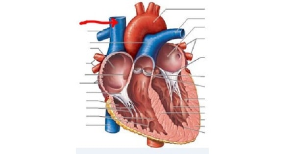
A. Opens into right ventricle
B. Enters the heart at level of 3rdcostal
C. Pierces paricardium at 2nd costal cartilage
D. Receives azygos vein behind sternal angle
Show Answer
|
Correct Answer » A Explanation |
 |
Ans;A. Opens into right ventricle
This structure is superior venacava
SVC opens in to right atrium (not right ventricle).
Superior vena-cava
- SVC collects blood from the upper half of the body and drains into the right atrium.
- It is formed by the union of two brachiocephalic veins at the level of lower border of 1″ right costal cartilage.
- It passes vertically downwards behind the right border of sternum and piercing the pericardium at the level of the second costal cartilage, enters the upper border of the right atrium to end in the upper and posterior part of sinus venorum at the lower border of third right costal cartilage.
Structure and Course of superior vena cava
- It is formed by the left and right brachiocephalic veins (also referred to as the innominate veins), which also receive blood from the upper limbs, eyes and neck, behind the lower border of the first right costal cartilage.
- It passes vertically downwards behind first intercostal space and receives azygos vein just before it pierces the fibrous pericardium opposite right second costal cartilage and its lower part is intrapericardial.
- And then, it ends in the upper and posterior part of the sinus venarum of the right atrium, at the upper right front portion of the heart.
- No valve divides the superior vena cava from the right atrium. As a result, the (right) atrial and (right) ventricular contractions are conducted up into the internal jugular vein and, through the sternocleidomastoid muscle, can be seen as the jugular venous pressure.
