


Question
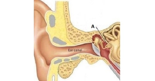
The structure marked as “A” in the image below develops from?

A. 1st arch.
B. 2nd arch.
C. 3rd arch.
D. 4th arch.

|
Correct Answer » B Explanation |
 |

Ans:B. Second Arch.
The structure marked as A in the picture above represents stapes.
Stapes develops from second arch except its footplate and annular ligament which are derived from the otic capsule.
Malleus and incus are derived from mesoderm of 1″ arch.
DERIVATIVES OF PHARYNGEAL ARCH
| Pharyngeal
arch |
Muscular contributions | Skeletal contributions | Nerve | Artery |
| 1st (also called “mandibular arch”) | Muscles of mastication,anterior belly of the digastric, mylohyoid,
tensor tympani, tensor veli palatini |
Premaxilla, maxilla,
mandible (only as a model for mandible not actual formation of mandible), zygomatic bone, part of the temporal bone, the incus, and the malleus of the middle ear, also Meckel’s cartilage and the sphenomandibular ligament. |
Trigeminal nerve(part of V2 and V3) | Maxillary artery, external carotid artery |
| 2nd (also called the “hyoid arch“) | Muscles of facial expression, buccinator,
platysma, stapedius,stylohyoid, posterior belly of the digastric, auricular |
Stapes, temporal styloid process, hyoid bone (lesser horns and upper part of body), stylohyoid ligament, Reichert’s cartilage | Facial nerve (VII) | Stapedial artery, hyoid artery |
| 3rd | Stylopharyngeus | Hyoid bone (greater horns and lower part of body), thymus | Glossopharyngeal nerve (IX) | common carotid, internal carotid |
| 4th | Cricothyroid muscle, all intrinsic muscles of soft palate (including levator veli palatini) except tensor veli palatini | Thyroid cartilage, epiglottic cartilage | Vagus nerve (X), superior laryngeal nerve | Right 4th aortic arch: subclavian artery
Left 4th aortic arch: aortic arch |
| 6th | All intrinsic muscles of larynx except the cricothyroid muscle | Cricoid cartilage, arytenoid cartilages, corniculate cartilage, cuneiform cartilages | Vagus nerve (X), recurrent laryngeal nerve | Right 6th aortic arch: pulmonary artery
Left 6th aortic arch: pulmonary artery and ductus arteriosus |