Question
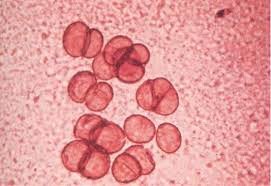
Identify the bodies seen in KOH mount of a case of Chromomycosis as shown in the photomicrograph below?
A. Cigar rosette bodies.
B. Sphagetti bodies.
C. Sclerotic bodies.
D. None of the above.
Show Answer
|
Correct Answer » C
Explanation
|
|
Ans: C. Sclerotic Bodies.
Chromoblastomycosis(Chromomycosis)
- It is a chronic fungal infection of the skin and the subcutaneous tissue caused by traumatic inoculation of a specific group of dematiaceous fungi.
- The infection usually results from a traumatic cutaneous injury that is often not remembered or realized by the patient. The agents often gain entry into the human body by contact with wood splinters or thorns.
- The lesions develop slowly at the site of implantation, producing a warty nodule, which tends to be limited to the skin and the subcutaneous tissue.
- Direct examination of 10% potassium hydroxide cleared lesion scrapings is by far the most useful. Typical, thick-walled, cigar-colored, sclerotic cells, also known as Medlar bodies, are readily seen, sometimes more colorfully referred to as golden-brown septate “copper pennies.