


Question
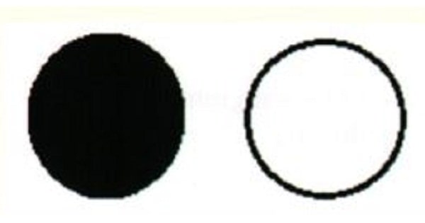
Visual defect shown in the photograph below occur in lesion of ?

A. Optic nerve.
B. Optic chiasma.
C. Geniculate body.
D. Visual cortex.
Show Answer


|
Correct Answer » A Explanation |
 |

Ans:A. Optic Nerve
Lesions of visual pathway
| No | Site of lesion | Causes | Features |
| 1 | – | – | Normal |
| 2 | Optic nerve | Optic neuritis
Optic atrophy Sarcoidosis LHON Traumatic avulsion |
Complete blindness in affected eye
Loss of light reflex on the affected side and consensual reflex loss on the other side Near reflex present Central/paracentral scotoma |
| 3 | Central chiasma | Pituitary tumors (MCC)
Suprasellar aneurysm, Craniopharyngioma, Glioma of Ill ventricle |
Bitemporal hemianopia
Bitemporal hemianopic paralysis of pupillary reflexes |
| 3a | Lateral chiasma | Distension of 3rd ventricle,
Atheroma of post. communicating A |
Binasal hemianopia
Binasal hemianopic paralysis of pupillary reflexes |
| 4 | Optic tract
Geniculate body |
Syphilitic meningitis, gumma, tuberculosis, tumors of optic thalamus | ncongruous (contralateral) homonymous hemianopia Contralateral Wernicke’s pupil (optic tract) |
| 5 | Optic radiations in temporal lobe (inferior fibres) | Superior quadrantic hemianopia (pie in the sky)
Normal pupillary reactions |
|
| 5a | Optic radiations in parietal lobe (Superior fibres) | Inferior quadrantic hemianopia (pie on the floor)
Normal pupillary reactions |
|
| 6 | Optic radiations | Homonymous hemianopia | |
| 7 | Anterior occipital (visual) cortex | Occlusion of posterior cerebral artery | Congruous homonymous hemianopia sparing macula |
| 8 | Tip of occipital cortex | Congruous homonymous macular defect |