


Question
Area as marked by an arrow is ?

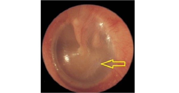
A. Shrapnell’s membrane.
B. Annulus tympanicus.
C. Pars tensa.
D. Posterior malleal fold.
Show Answer
|
Correct Answer » C Explanation |
 |

Ans:C. Pars tensa.
The image is shown of a Tympanic membrane.
Tympanic membrane
- Structure:
- Outer epithelial/cutaneous layer: stratified squamous epithelium .
- Middle fibrous layer: thin/absent in pars flaccida; contains 3 types of fibers – radial,
- circular and parabolic.
- Inner mucosal layer
Components:
- Anterior and posterior malleolar fold: Converges to the lateral process of malleus from 2 ends of the tympanic notch of Rivinus.
- Pars flacidda/Sharpnell membrane/Rivinus membrane: Part of relatively lax membrane lying above the malleolar folds; lack middle fibrous layer
- Pars tensa: Relatively taut and forms the remaining part of the tympanic membrane; contain all 3 layers.
- Annulus tympanicus: Thick rolled periphery of pars tensa.
- Umbo: Center of the tympanic membrane which divides it into 4 quadrants and the point of attachment for the handle of malleus.
- Tympanic notch of Rivinus/Rivinus incisure: Notch (deficient area) in the superior part of fibrocartilaginous tympanic ring bridged by pars flaccida.
- Cone of light/Light reflex: In the antero-inferior quandrant, below and anterior to the handle of malleus.