

Question

A. Pneumatocoele.
B. Annular pancreas.
C. Jejunal atresia.
D. Intestinal Perforation.
|
Correct Answer » A Explanation |
|

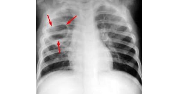
Inflammatory pneumatocoeles result from a rapidly progressive inflammation in which there are plugging of the smaller bronchi in the affected areas, destruction of the distal alveolar tissue, and cystic hyperexpansion of the air space. Pneumatocoeles are found much more often in children than in adults.
They are seen particularly in infants and children with staphylococcal pneumonia.
However, they have also been described in pneumonia due to other micro-organisms such as Streptococcus pneumoniae, Klebsiella, Hemophilus influenzae, and in measles pneumonitis.
On chest radiographs, they appear as thin-walled,rounded radiolucencies in areas of pneumonic consolidation. They can be single or multiple.
As the surrounding consolidation resolves, the pneumatocoeles become more apparent and/or enlarge. Thus, they are usually seen during the resolution stage of pneumonitis.
Most often, however, they resolve slowly and disappear within two to three weeks