
Question
Diagnose the fracture shown in the picture below ?
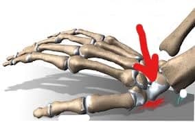
A. Fracture Scaphoid.
B. Fracture Colles.
C. Fracture Lunate.
D. Facture Olecranon.

|
Correct Answer » A Explanation |
 |
The scaphoid is one of eight small bones that make up the “carpal bones” of the wrist. It connects two rows of these bones – the proximal row (closer to the forearm) and the distal row (closer to the hand). This connection puts it at extra risk for injury.
A fracture of the scaphoid bone usually occurs from a fall onto the outstretched hand.
Pain (with or without swelling or bruising at the “thumb side” of the wrist) can be noticed within days following a fall. Because there is no visible deformity and no difficulty with motion, many people with this injury assume that it is a wrist “sprain.” Unfortunately, delaying treatment can cause problems. Visit a hand surgeon as soon as possible if you think you might have fractured your wrist.
Diagnosis
Scaphoid fractures are usually diagnosed by an x-ray of the wrist; however, x-rays do not always show scaphoid fractures. A break in the bone that cannot be seen on x-ray yet is called an “occult” fracture. If you are tender directly over the scaphoid bone which is located in the hollow at the thumb side of the wrist.
A CT scan, bone scan or MRI may also be used to diagnosis the fracture.
TREATMENT
If the fracture is non-displaced (bone has not moved out of place at the fracture), it usually can be successfully treated with a cast. Although the fracture may heal in as little as six weeks, it may take longer for some patients.
If the fracture is in a certain part of the bone or if the fracture is at all displaced (bone ends have shifted), surgery might be the best option. This might include the insertion of a screw or pins.
