|
Correct Answer » D
Explanation
|
|
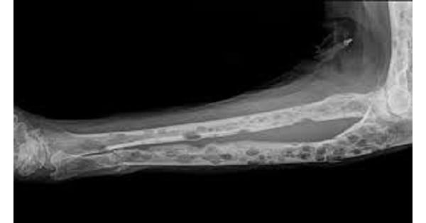
The condition shown in the picture above represents Multiple myeloma (Appearance: Multiple punched out lesions).
Multiple myeloma is characteristically associated with Plasmacytosis >10%.
Multiple myeloma may be associated with both Hypogammaglobulinemia and Hypergammaglobulinemia.
Electrophoresis Pattern in Multiple Myeloma
Monoclonal Hypergammaglobulinemia (M spike) This is the characteristic electrophoresis pattern in Myeloma
Hypogammaglobulinemia
- About 10-20% of multiple myeloma patients present with Hypogammaglobulinemia on electrophoresis.
- This configuration is suggestive of light chain variant of multiple myeloma in which Bence Jones proteins are excreted into urine without a serum myeloma protein being evident with routine electrophoresis techniques.
`Multiple Myeloma results in hypergammaglobulinemia of a specific class of immunoglobulins.
Hypogammaglobulinemia also justifies a search for myeloma especially for the possibility of light chain disease. ‘
Multiple Myeloma may be with Bence Jones Proteins, Amyloidosis & Renal failure.