Question
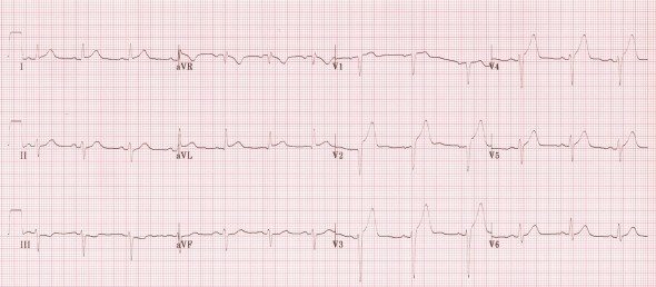
Blockage in which blood vessel would have resulted in the condition shown by ECG below:
A. Left Anterior Descending Artery.
B. Left Coronary Artery.
C. Right Coronary Artery.
D. Right Circumflex artery.
Show Answer
|
Correct Answer » A
Explanation
|
|
Ans: A. Left Anterior Descending Artery
ECG in the image shows:
Hyperacute Anteroseptal STEMI
- ST elevation is maximal in the anteroseptal leads (V1-4).
- Q waves are present in the septal leads (V1-2).
- There is also some subtle STE in I, aVL and V5, with reciprocal ST depression in lead III.
- There are hyperacute (peaked ) T waves in V2-4.
Anterior STEMI
- It results from occlusion of the left anterior descending artery (LAD).
- Anterior myocardial infarction carries the worst prognosis of all infarct locations, mostly due to larger infarct size.
ECG findings:
- ST segment elevation with Q wave formation in the precordial leads (V1-6) ± the high lateral leads (I and aVL).
- Reciprocal ST depression in the inferior leads (mainly III and aVF).
The different infarct patterns are named according to the leads with maximal ST elevation:
- Septal = V1-2
- Anterior = V2-5
- Anteroseptal = V1-4
- Anterolateral = V3-6, I + aVL
- Extensive anterior / anterolateral = V1-6, I + aVL