
Question
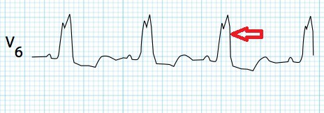
Diagnose the condition

A. Complete Heart Block.
B. Right bundle branch block.
C. Left Bundle Branch Block.
D. 2nd degree heart block(Type 2).
Show Answer

|
Correct Answer » C Explanation |
 |
Ans: C. Left Bundle branch block.
ECG in the image shows:Dominant S wave(‘W’ shaped) in V1 with broad, notched (‘M’-shaped) R wave in V6
Left Bundle branch Block
- Normally the septum is activated from left to right, producing small Q waves in the lateral leads.
- In LBBB, the normal direction of septal depolarisation is reversed (becomes right to left), as the impulse spreads first to the RV via the right bundle branch and then to the LV via the septum.This sequence of activation extends the QRS duration to > 120 ms and eliminates the normal septal Q waves in the lateral leads.
- The overall direction of depolarisation (from right to left) produces tall R waves in the lateral leads (I, V5-6) and deep S waves in the right precordial leads (V1-3), and usually leads to left axis deviation.
- As the ventricles are activated sequentially (right, then left) rather than simultaneously, this produces a broad or notched (‘M’-shaped) R wave in the lateral leads.
ECG criteria:
- QRS duration of > 120 ms
- Dominant S wave in V1
- Broad monophasic R wave in lateral leads (I, aVL, V5-V6)
- Absence of Q waves in lateral leads (I, V5-V6; small Q waves are still allowed in aVL)
- Prolonged R wave peak time > 60ms in left precordial leads (V5-6)
- Left axis deviation
Causes
- Aortic stenosis
- Dilated cardiomyopathy
- Acute myocardial infarction
- Extensive coronary artery disease
- Primary disease of the cardiac electrical conduction system
- Long standing hypertension leading to aortic root dilatation and subsequent aortic regurgitation
- Lyme disease
Treatment
- Those with LBBB and syncope or near-syncope may require a pacemaker.
