Question
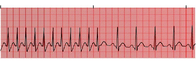
A patient presented with palpitations.ECG showed the following picture.What can be the most probable diagnosis?
A. Unifocal Atrial Tachycardia.
B. Multifocal Atrial Tachycardia.
C. Ventricular Tachycardia.
D. Atrial Fibrillation.
Show Answer
|
Correct Answer » A
Explanation
|
|
Ans: A. Unifocal Atrial Tachycardia.
Atrial Tachycardia(Paroxysmal Atrial Tachycardia ,unifocal atrial tachycardia, ectopic atrial tachycardia)
- Atrial tachycardia is a form of supraventricular tachycardia, originating within the atria but outside of the sinus node.
Pathophysiology of Atrial Tachycardia
- Usually due to single ectopic focus.
- The underlying mechanism can involve reentry, triggered activity or increased automaticity.
- May be paroxysmal or sustained.
- Multiple causes including digoxin toxicity, atrial scarring, catecholamine excess, congenital abnormalities; may be idiopathic.
- Sustained atrial tachycardia may rarely be seen and can progress to tachycardia-induced cardiomyopathy.
ECG Features of Atrial Tachycardia
- Atrial rate > 100 bpm.
- P wave morphology is abnormal when compared with sinus P wave due to ectopic origin.
- There is usually an abnormal P-wave axis (e.g. inverted in the inferior leads II, III and aVF)
- At least three consecutive identical ectopic p waves.
- QRS complexes usually normal morphology unless pre-existing bundle branch block, accessory pathway, or rate related aberrant conduction.
- Isoelectric baseline (unlike atrial flutter).
- AV block may be present — this is generally a physiological response to the rapid atrial rate, except in the case of digoxin toxicity where there is actually AV node suppression due to the vagotonic effects of digoxin, resulting in a slow ventricular rate (“PAT with block”).