


Question
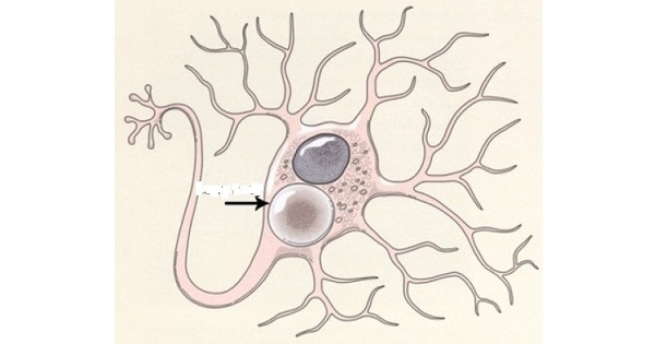
Body shown in the photomicrograph below is found in ?


A. Alzeihmer’s disease.
B. Astrocytoma.
C. Parkinson’s disease
D. Huntington’s disease.
Show Answer


|
Correct Answer » C Explanation |
 |

Ans:C. Parkinson’s disease
Body shown in the photomicrograph above represents Lewy bodies.
Lewy bodies
- They are abnormal aggregates of protein that develop inside nerve cells in Parkinson’s disease (PD), Lewy body dementia, and some other disorders.
- They are identified under the microscope when histology is performed on the brain.
- A classical Lewy body is an eosinophilic cytoplasmic inclusion consisting of a dense core surrounded by a halo of 10-nm-wide radiating fibrils, the primary structural component of which is alpha-synuclein.