
Question
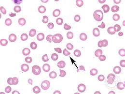
Cell type (Arrows)shown in photograph are found in
A. HUS
B. TTP
C. DIC
D. All of the above
Show Answer

|
Correct Answer » D Explanation |
 |
Ans: D. All of the above
The image is showing Schistocyte in the blood smear film.
Schistocyte
- It is a fragmented part of a red blood cell.
- Schistocytes are typically irregularly shaped, jagged, and have two pointed ends.
- A true schistocyte does not have central pallor.Schistocytes are sometimes referred to as “helmet cells”.
Causes:
- Several microangiopathic diseases, including disseminated intravascular coagulation and thrombotic microangiopathies, generate fibrin strands that sever red blood cells as they try to move past a thrombus, creating schistocytes.
- Schistocytes are often seen in patients with hemolytic anemia. They are frequently a consequence of mechanical artificial heart valves and hemolytic uremic syndrome, thrombotic thrombocytopenic purpura.
- Excessive schistocytes present in blood can be a sign of microangiopathic hemolytic anemia .
