
Question
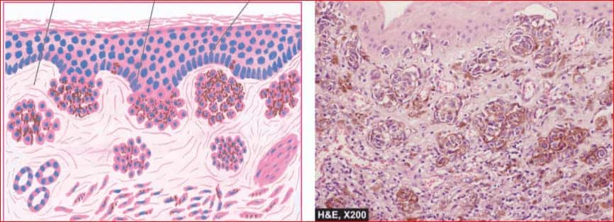
A cut section of Dermis and Epidermis is shown in the image.Identify the pigment deposited.
A. Hemosiderin
B. Porphyrin
C. Melanin
D. Lipofuscin
Show Answer

|
Correct Answer » C Explanation |
 |
Ans:C. Melanin.
Image shows:Compound naevus showing clusters of benign naevus cells in the dermis as well as in lower epidermis. These cells contain coarse, granular, brown-black melanin pigment.
ENDOGENOUS PIGMENTS
- Melanin
- Melanin-like pigment a. Alkaptonuria b. Dubin-Johnson syndrome
- Haemoprotein-derived pigments i) Haemosiderin ii) Acid haematin (Haemozoin) c. Bilirubin d. Porphyrins
- Lipofuscin (Wear and tear pigment).
MELANIN
- Melanin is the brown-black, non-haemoglobin-derived pigment normally present in the hair, skin, choroid of the eye, meninges and adrenal medulla.
- It is synthesised in the melanocytes and dendritic cells, both of which are present in the basal cells of the epidermis and is stored in the form of cytoplasmic granules in the phagocytic cells called the melanophores, present in the underlying dermis.
- Melanocytes possess the enzyme tyrosinase necessary for synthesis of melanin from tyrosine.
- The presence of tyrosinase can be detected by incubation of tissue section in the solution of dihydroxy phenyl alanine (DOPA). If the enzyme is present, dark pigment is identified in pigment cells. This test is called as DOPA reaction.
Various disorders of melanin pigmentation cause generalised and localised hyperpigmentation and hypopigmentation:
i) Generalised hyperpigmentation:
- In Addison’s disease
- Chloasma
- In chronic arsenical poisoning
ii) Focal hyperpigmentation:
- Cäfe-au-lait spots are pigmented patches seen in neurofibromatosis and Albright’s syndrome.
- Peutz-Jeghers syndrome is characterised by focal peri-oral pigmentation.
- Melanosis coli is pigmentation of the mucosa of the colon.
- Melanotic tumours, both benign such as pigmented naevi , and malignant such as melanoma.
- Lentigo is a pre-malignant condition in which there is focal hyperpigmentation on the skin of hands, face, neck, and arms.
- Dermatopathic lymphadenitis is an example of deposition of melanin pigment in macrophages of the lymph nodes draining skin lesions.
iii) Generalised hypopigmentation:
- Albinism is an extreme degree of generalised hypopigmentation in which tyrosinase activity of the melanocytes is genetically defective .
iv) Localised hypopigmentation:
- Leucoderma is a form of partial albinism
- Vitiligo.
- Acquired focal hypopigmentation can result from various causes such as leprosy, healing of wounds, DLE, radiation dermatitis etc.
