
Question
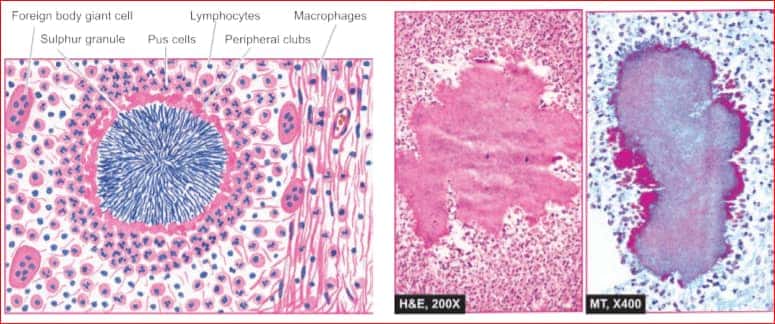
A patient presented with a jaw swelling.Histopathological examination shows the following picture.At which other places can this type of infection occur in the body?
A. Thoracic
B. Abdominal
C. Pelvic
D. All of the above
Show Answer

|
Correct Answer » D Explanation |
 |
Ans:D. All of the above.
The patient in question is suffering from Actinomycosis.
Image shows:Actinomycosis. Microscopic appearance of sulphur granule lying inside an abscess. The margin of the colony shows hyaline filaments highlighted by Masson’s trichrome stain (right photomicrograph).
ACTINOMYCOSIS
- Actinomycosis is a chronic suppurative disease caused by anaerobic bacteria, Actinomycetes israelii.
- The organisms are commensals in the oral cavity, alimentary tract and vagina. The infection is always endogeneous in origin and not by person-to-person contact.
- The organisms invade, proliferate and disseminate in favourable conditions like break in mucocutaneous continuity, some underlying disease etc.
MORPHOLOGIC FEATURES.
- Depending upon the anatomic location of lesions, actinomycosis is of 4 types: cervicofacial, thoracic, abdominal, and pelvic .
- Microscopically, the following features are seen :
- i) The inflammatory reaction is a granuloma with central suppuration. There is formation of abscesses in the centre of lesions and at the periphery chronic inflammatory cells, giant cells and fibroblasts are seen.
- ii) The centre of each abscess contains the bacterial colony, ‘sulphur granule’, characterised by radiating filaments (hence previously known as ray fungus) with hyaline, eosinophilic, club-like ends representative of secreted immunoglobulins.
- iii) Bacterial stains reveal the organisms as gram-positive filaments, nonacid-fast, which stain positively with Gomori’s methenamine silver (GMS) staining.
