
Question
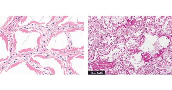
A newborn infant begins with dyspnoea within a few hours after birth with tachypnoea, hypoxia and cyanosis.Histopathological examination of lung shows the following picture.What can be a probable cause for this condition?
A. Preterm infant
B. Infant born to diabetic mother
C. Delivery by caesarean section
D. All of the above.

|
Correct Answer » D Explanation |
 |
Ans:D. All of the above.
Image shows:Histological appearance of hyaline membrane disease. There are alternate areas of collapsed and dilated alveolar spaces, many of which are lined by eosinophilic hyaline membranes.
The patient is suffering from Hyaline Membrane Disease(Acute Respiratory Distress Syndrome:ARDS).
ACUTE RESPIRATORY DISTRESS SYNDROME (HYALINE MEMBRANE DISEASE)
ETIOLOGY.
Neonatal ARDS is primarily initiated by hypoxia, either shortly before birth or immediately afterward. It occurs in following clinical settings:
- Preterm infants
- Infants born to diabetic mothers
- Delivery by caesarean section
- Infants born to mothers with previous premature infants
- Excessive sedation of the mother causing depression in respiration of the infant
- Birth asphyxia from various causes such as coils of umbilical cord around the neck
- Male preponderance (1.5 to 2 times) over female babies due to early maturation of female lungs
Adult ARDS may occur from the following causes:
- Shock due to sepsis, trauma, burns
- Diffuse pulmonary infections, chiefly viral pneumonia
- Pancreatitis
- Oxygen toxicity
- Inhalation of toxins and irritants e.g. smoke, war gases, nitrogen dioxide, metal fumes etc.
- Narcotic overdose
- Drugs e.g. salicylates, colchicine
- Aspiration pneumonitis
- Fat embolism
- Radiation
MORPHOLOGIC FEATURES.
Grossly, the lungs are normal in size.
They are characteristically stiff, congested, heavy and airless so that they sink in water.
Microscopically, the important features are as follows:
- There is presence of collapsed alveoli (atelectasis) alternating with dilated alveoli.
- Necrosis of alveolar epithelial cells and formation of characteristic eosinophilic hyaline membranes lining the respiratory bronchioles, alveolar ducts and the proximal alveoli. The membrane is largely composed of fibrin admixed with cell debris derived from necrotic alveolar cells.
- Interstitial and intra-alveolar oedema, congestion and intra-alveolar haemorrhages.
- Changes of bronchopneumonia may supervene.
- With time, compensatory proliferation of pneumocytes into alveolar lumen may be seen as tufts of alveolar epithelium.
- In organising stage, there is interstitial fibrosis obliterating alveolar spaces.
