|
Correct Answer » B
Explanation
|
|
Ans:B. Rarely involute spontaneously
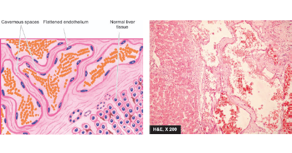
Image shows: Cavernous haemangioma of the liver. The vascular spaces are large, dilated, many containing blood, and are lined by flattened endothelial cells. Scanty connective tissue stroma is seen between the cavernous spaces.
CAVERNOUS HAEMANGIOMA.
- Cavernous haemangiomas are single or multiple, discrete or diffuse, red to blue, soft and spongy masses.
- They are often 1 to 2 cm in diameter.
- They are most common in the skin (especially of the face and neck); other sites are mucosa of the oral cavity, stomach and small intestine, and internal visceral organs like the liver and spleen.
- Histologically, cavernous haemangiomas are composed of thin-walled cavernous vascular spaces, filled partly or completely with blood.
- The vascular spaces are lined by flattened endothelial cells.
- They are separated by scanty connective tissue stroma .
- Cavernous haemangiomas rarely involute spontaneously.