


Question
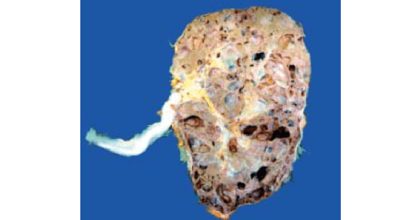
The image shows presence of :

A. Medullary Sponge Kidney
B. Renal Cystic Dysplasia
C. Autosomal dominant polycystic kidney disease
D. Autosomal recessive polycystic kidney disease
Show Answer


|
Correct Answer » C Explanation |
 |

Ans:C. Autosomal dominant polycystic kidney disease.
Image shows:Adult (autosomal dominant) polycystic kidney disease(ADPKD). The kidney is enlarged and heavy. Sectioned surface shows loss of demarcation between cortex and medulla and replacement of the
entire renal parenchyma by cysts varying in diameter from a few millimeters to 4-5 cm. These cysts are not communicating with the pelvicalyceal system. The renal pelvis and calyces are distorted due to cystic change.
CYSTIC DISEASES OF KIDNEY

