


Question
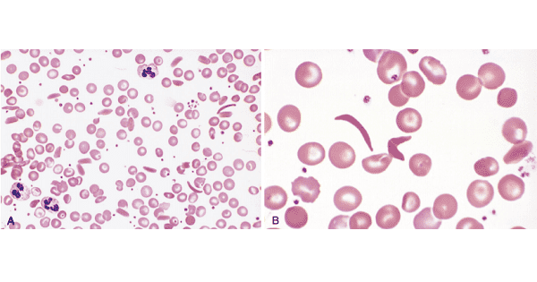
A patient is suffering from anemia.Peripheral blood smear is shown in the image.Regarding this pathology,which of the following statements is false?

A. Vaso-occlusive crisis or Pain crisis is the most common clinical manifestation
B. Intravascular hemolysis
C. Autosomal recessive disease
D. Caused by a point mutation.


|
Correct Answer » B Explanation |
 |

Ans:B. Intravascular hemolysis.
The patient in question is suffering from Sickle cell anemia.Extravascular hemolysis is seen in this condition.
Image shows:Sickle cell anemia—peripheral blood smear. A, Low magnification shows sickle cells, anisocytosis, poikilocytosis, and target cells. B, Higher magnification shows an irreversibly sickled cell in the center.
Image shows:
SICKLE CELL DISEASE
- Normal adult red cells contain mainly HbA(α2β2), along with small amounts of HbA2(αδ2) and fetal hemoglobin HbF(α2γ2)
- Sickle cell disease is caused by a point mutation(missense mutation) at the 6th codon of β globin chain leading to substitution of glutamine by valine.
- Sickle cell disease is inherited as Autosomal recessive.
- Sickle cell anemia is the clinical manifestation of homozygous genes
- Sickle cell trait (heterozygotes) gives protection against falciparum malaria
- In sickle cell trait 40% is HbS and rest is HbA.
- Sickle cell anaemia is commonly seen in black
PATHOGENESIS OF SICKLE CELL DISEASE
- Sickle cell disease causes extravascular hemolysis.
- Sticky patch in sickle cell disease results from replacement of a polar residue (Glutamate) with a non polar residue (Valine) at the 6th position of the beta chain.
- RFLP results from a single base change in Sickle cell disease.
- Decreased Solubility is the cause of sickling of RBC in sickle cell disease.
- Deoxygenated HbS undergo aggregation and polymerization, producing a distorted sickle or holly-leaf shaped RBC which block microcirculation.
- Initial sickling is reversible on oxygenation, but after repeated aggregation, sickling is irreversible.
- There is a positive correlation between HBS and polymerization of HBS in Sickle cell anemia.
- HbF inhibit polymerization of HbS; hence infants are asymptomatic until 6 months
- In hereditary persistence of HbF – the disease is less severe
- HbC (lysine is substituted for glutamate in the 6th amino acid residue of β globin) has greater tendency to form aggregates than HbS – but disease is milder in HbSC
- Factors precipitating sickling
- Dehydration (↑MCHC)
- Hypoxia
- Acidosis (↓ pH)
- Increased transit time of red cells through microvascular beds
- Infections
CLINICAL FEATURES OF SICKLE CELL DISEASE
- Vaso-occlusive crisis or Pain crisis (hall mark) – most common clinical manifestation
- Bones (especially the back and long bones) and the chest are commonly affected
- Plugging of small vessels in bones – dactylitis (hand and foot syndrome)
- Avascular necrosis of femoral head, Osteomyelitis due to Salmonella.
- Reccurent infections is the most common cause of death in Sickle cell anemia.
- Acute chest syndrome
- Chest pain, tachypnea, fever, cough, and arterial oxygen desaturation
- Pulmonary infarction and pneumonia are the most frequent underlying conditions
- Extramedullary hematopoiesis prominent cheek bones and crew-hair cut appearance of skull
- Occlusion of vertebral arteries – fish mouth deformity of vertebrae
- Sequestration crisis – massive sequestration of sickled red cells leads to rapid splenic enlargement,hypovolemia and shock.
- In chronic disease due to repetititve infarction, there is progressive shrinkage of spleen following repeated episodes of sequestration crisis – autosplenectomy
- Children are more prone for Pneumococcus pneumoniae and H influenzae septicemia and meningitis with capsulated organisms like pneumococci
- Aplastic crisis – transient cessation of marrow erythropoiesis due to an acute infection with parvovirus B19.
- Unlike other crisis reticulocyte count is low
- Stroke is especially common in children and less common in adults
- Priapism may result in permanent impotence
- Increased break down of hemoglobin can cause pigment gallstones and hyperbilirubinemia.
- Renal papillary necrosis leads to hyposthenuria (inability to concentrate urine) and gross hematuria (less common in sickle cell disease)
- Leg ulcers, delayed puberty, pulmonary hypertension, retinopathy
- Granulocytosis is common
- PAH,Cardiomegaly
- Pulmonary hypertension.
- ‘Sea-Fan’ retina is seen.
- Decreased Osmotic Fragility
- Sickle cell anemia is a non-metabolic cause of abdominal pain.
- Female gender: less severe manifestations since HbF is high.
DIAGNOSIS OF SICKLE CELL DISEASE
- Peripheral blood: sickled cells, target cells, Howell-Jolly bodies, Anemia (extravascular hemolysis), reticulocytosis
- ↓ESR (raised in other anemias)
- Metabisulfite and sodium dithionite which consumes oxygen in a sample of blood, precipitates sickling if HbS is present – useful screening test
- Definite diagnosis by – Hemoglobin electrophoresis (which reveal 60% HbS),HbA2 is normal; HbF in varying concentrations: HbA absent
- Alkali denaturation method – to determine concentration of HbF (HbF is relatively resistant to denaturation by strong alkali)
- Quantitative estimation of HbF – Kleihauer test.
- Polymerase chain reaction with allele-specific oligonucleotide hybridization, Southern blot analysis ,DNA sequencing can be used to detect the point mutation in the beta globin gene that causes sickle cell anemia.
- Gandy gamma bodies are seen in sickle-cell anaemia .
- Vertebra plana ,Bone infarct , Marrow hyperplasia,’H’ shaped vertebra are the radiological features of Sickle cell anemia.