


Question
A 72-year-old woman with a history of poorly controlled hyperlipidaemia, hypertension and diabetes mellitus suddenly lost consciousness and developed left-sided weakness.
Physical examination showed a BP of 210/120. An urgent CT brain was performed.What is the likely diagnosis?

A. Subdural hematoma
B. Subarachnoid Hemorrhage
C. Extradural Hematoma
D. Cerebral Infarct
Show Answer


|
Correct Answer » D Explanation |
 |

Ans:D. Cerebral Infarct.
Image shows:
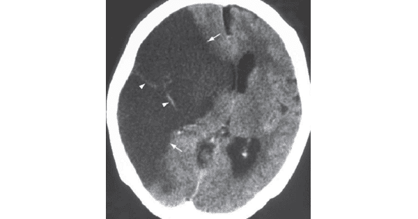
- Non-contrast axial CT scan showing a wedge shaped area of low attenuation (arrows), with effacement of underlying cortical sulci, involving both grey and white matter in the right MCA territory.
- Also note the mass effect on the ventricles and contralateral shift. The diagnosis is an acute right MCA territory infarct.
- The arrowheads show a hyperdense thrombus in the branches of the right MCA.
Cerebral Infarct
- Cerebral infarction is the irreversible loss of brain tissues due to ischaemia or haemorrhage
- CT is the initial imaging of choice for acute stroke. However, it may not demonstrate a cerebral infarct during the first 12 hours.
- At this time its role is to rule out the presence of intracranial haemorrhage as this defines the treatment options.
- An infarct may be treated by anticoagulants/ thrombolytics whereas these are contraindicated in the presence of an intracranial haemorrhage.
- Early signs of cerebral infarction on CT:
- Loss of grey-white matter interface
- Sulcal effacement
- Hyperdense clot within thrombosed artery (dense MCA sign ).