
Question
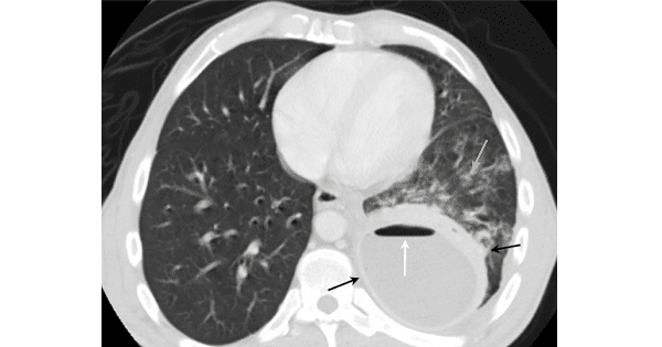
A 67 year old patient presented with cough.CT scan thorax shows the following features.What can be the most possible diagnosis?
A. Hiatal Hernia
B. TB
C. Lung Abscess
D. Sarcoidosis

|
Correct Answer » C Explanation |
 |
Ans:C. Lung Abscess.
Image shows:A single, contrast-enhanced axial CT scan image of the chest shows a large cavitary lesion in the left lower lobe with a relatively thick wall(black arrows). The cavity has a smooth inner margin and an air-fluid level (white arrow). There is inflammatory reaction in the surrounding lung (yellow arrow).
Lung Abscess
They may be consequences of aspiration (most commonly) or pneumonia or they may be secondary to bronchial obstruction, bronchiectasis, bacterial endocarditis or spread of infection from elsewhere in the lung
Most abscesses are caused by mouth anaerobes in patients with pre-existing periodontal disease
Alcoholics (most commonly), those with seizure disorders leading to aspiration and those with poor dentition are at a higher risk
Anaerobes are most frequent cause; most lung abscesses are polymicrobial in etiology
- Peptostreptococcus
- Bacteroides
- Fusobacterium
Also
- Staphylococcus
- Streptococcus pyogenes
- Klebsiella
- Haemophilus influenzae
- Actinomyces species
Clinical Findings
- Typically take 7-14 days from pneumonia to abscess formation, depending on agent
- Fever
- Cough
- Foul-smelling sputum production
- Hemoptysis
- Night sweats
- Anorexia and weight loss
Imaging Findings
- On conventional radiography
- Usually single cavity
- Cavities typically have a
- Thick-wall (which may become thinner as the surrounding inflammation resolves)
- Smooth inner margin
- Air-fluid level
- More frequent in superior segments of lower lobes or posterior segments of lower lobes
- Unlike pleural collections, lung abscesses frequently have a fluid level which is approximately the same length on both the frontal and lateral projection
- About 1/3 may have an associated empyema
CT
- Helpful in differentiating between a lung abscess and an empyema
- Cavity may be seen as rounded with a thick wall and an air-fluid level
- Lung abscesses, being intraparenchymal, form an acute angle where they meet the chest wall
Differential Diagnosis
- Cavitary bronchogenic carcinoma
- TB
- Multiple cavities point toward a necrotizing pneumonia more than lung abscess
- Multiple cavitating masses point towards septic emboli
Treatment
- Antibiotics for 4-6 weeks or until the chest radiograph shows resolution
- Percutaneous catheter drainage
- Surgery, either a lobectomy or pneumonectomy, may be used for patients who fail to respond to medical therapy
Complications:
- Empyema
- Bronchopleural fistula
- Fibrosis of pleura
