


Question
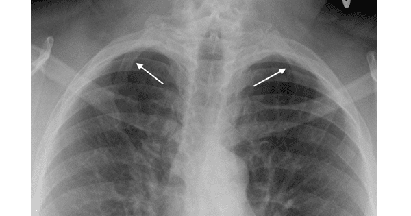
A patient presents with pain in his upper extremities and tingling and numbness in his 4th and 5th digits of his hands.X ray shows the following features.Which of the following structures is most likely being compressed?

A. Axillary artery
B. Brachial artery
C. Brachiocephalic artery
D. Subclavian artery
Show Answer


|
Correct Answer » D Explanation |
 |

Ans:D. Subclavian artery.
Image shows:Bilateral cervical ribs. There are bilateral accessory ribs arising from the 7th cervical vertebral body (white arrows).
CERVICAL RIB
- The rib or ribs arise from the 7th cervical vertebral body
- They are usually bilateral (80%)
- More common in females
- The anterior end of the cervical rib may attach to the first rib, the sternum or the cartilage of the first rib; it may be a fibrous band or it may have no anterior attachment
Clinical Findings
- Most are asymptomatic
- Less than 10% of those with cervical ribs will be symptomatic
- The presence of a cervical rib can cause a form of thoracic outlet syndrome due to compression of the lower trunk of the brachial plexus or subclavian artery. These structures become encroached upon by the cervical rib and scalene muscles.
- Compression of the brachial plexus may be identified by weakness of the muscles around the muscles in the hand, near the base of the thumb.
- Compression of the subclavian artery is often diagnosed by finding a positive Adson’s sign on examination, where the radial pulse in the arm is lost during abduction and external rotation of the shoulder.
Treatment
- Most patients improve with exercise and physical therapy
- Surgical intervention is unusual
- Indications for surgical resection of the accessory rib may include disabling pain, paresthesias and failure of conservative treatment