Question
A 70 year old male patient presented with headache.Xray showed the following features.What can be the most possible diagnosis?
A. Multiple Myeloma
B. Hyperparathyroidism
C. Paget’s Disease
D. Osteopetrosis
Show Answer

|
Correct Answer » C Explanation |
 |
Ans:C. Paget’s Disease.
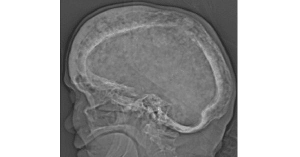
Image shows:Paget involvement of the skull, with widening of the diploic space, typical “cotton wool” appearance and over-riding enlarged frontal bone (Tam O’Shanter sign).
PAGET’S DISEASE OF BONE (OSTEITIS DEFORMANS)
- Paget’s disease is characterized by increased bone turnover and enlargement and thickening of the bone, but the internal architecture is abnormal and the bone is usually brittle. Primary defect is in osteoclasts with increased osteoclastic activity. This results secondarily increase in osteoblastic activity (normal osteoclasts and osteoblasts act in a co-ordinated manner). So, characteristic cellular change is a marked increase in osteoclastic and osteoblastic activity. Bone turnover is acclerated, plasma alkaline phosphatase is raised (a sign of osteoblastic activity) and there is increased excretion of hydroxyproline in urine (due to osteoclastic activity).
- Viral infection (paramyxovirus) in association with genetic susceptibility has been postulated.
Clinical features of Paget’s disease
- Paget’s disease is slightly more common in males and is seen after 40 years of age.
- The pelvis and tibia being the commonest sites, and femur, skull, spine (vertebrae) and clavicle the next commonest .
- When patients does present, they present because any of the three : –
- Bone pain – usually the first symptom
- Kyphosis, bowed tibias, large head
- Frequent “chalkstick” fractures with slight trauma.
- Histology: Mosaic like appearance of osteoid secondary to rapid disordered bone resorption and production.
- Serum calcium and phosphate: normal.
- Alkaline phosphatase, Urinary hydroxyproline, Serum C-telopeptide (CTx): elevated.
Complications of Paget’s disease
- Fracture : Are common in weight bearing bones
- Cranial nerve compression : – May cause impaired vision, facial palsy, trigeminal neuralgia or deafness.
- Otosclerosis : – Another cause of deafness in Paget’s disease.
- Spinal canal stenosis and nerve root compression
- High output cardiac failure
- Osteoarthritis : of Hip and knee
- Rarely osteosarcoma
Imaging Findings
Classical triad
- Thickening of the cortex
- Accentuation of the trabecular pattern
- Increased size of bone
Cyst-like areas
Skull (involvement in 29-65%)
- Inner and outer table involved
- Leads to diploic widening
- Osteoporosis circumscripta is well-defined lysis, most commonly in frontal bone producing well-defined geographic lytic lesion in skull
- Represents early destructive phase of disease active stage)
- “Cotton wool” appearance represents mixed lytic and blastic pattern of thickened calvarium (later stage)
- Basilar invagination with encroachment on foramen magnum
- Deossification and sclerosis in maxilla
- Sclerosis of skull base
Long bones (almost invariably starts at end of bone)
- “Candle flame” or “blade of grass” pattern of lysis is the advancing tip of V-shaped lytic defect in diaphysis of long bone originating in subarticular site
- Lateral curvature of femur
- Anterior curvature of tibia (commonly resulting in fracture)
Pelvis
- Thickened trabeculae in sacrum, ilium
- Rarefaction in central portion of ilium (looks like a large lytic lesion)
- Thickening of iliopectineal line
- Acetabular protrusion with secondary degenerative joint disease
Spine (upper cervical, low dorsal, midlumbar most common sites)
- Coarse trabeculations at periphery of bone
- “Picture-frame vertebra” mimics bone-within-bone appearance
- Enlarged vertebral body with reinforced peripheral trabeculae and more lucent center, typically in lumbar spine
- “Ivory vertebra” is a blastic vertebra with increased density
- Ossification of spinal ligaments, paravertebral soft tissue, disk spaces can occur.
Management:
- Symptomatic patients are treated with bisphosphonates (e.g. Alendronate) aiming to reduce the bone turnover, to promote healing of osteolytic lesions and improve bone pain.