


Question
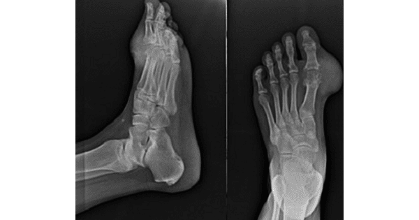
A 40 year old male presented with swelling of the great toe of left foot.X ray showed the following features.What can be the most possible diagnosis?

A. Rheumatoid Arthritis
B. Tuberculous Arthritis
C. Sarcoid
D. Gout.
Show Answer


|
Correct Answer » D Explanation |
 |

Ans:D. Gout.
Image shows:
X ray shows juxta-articular erosive changes around the first metatarsophalangeal(MTP) joint with overhanging edges and associated with a moderate soft tissue swelling.However the joint space is maintained.
No evidence of osteopenia.
GOUT
- Gout is a crystal arthropathy due to deposition of monosodium urate (MSU) crystals in and around the joints.
Epidemiology
- Typically occurs in those above 40 years. There is a strong male predilection of 20:1.
Pathology
- Characterised by monosodium urate crystals deposition in periarticular soft tissues. The crystals are needle-shaped and are strongly birefringent in plane polarised light .
- The synovial fluid is generally a poor solvent for monosodium urate and therefore causes crystallisation at low temperatures. The crystals are chemotactic and activate complement.
- Primary gout is a disorder of purine metabolism. Purine degradation result in the production of uric acid.
- Gout occur mainly from hyperuricemia secondary to reduced uric acid excretion from the kidney, and in a minority of cases it occur secondary to overproduction of uric acid.
- The first episode of acute gouty arthritis frequently begins at night with dramatic joint pain, swelling, warmth, redness, and tenderness.
- Usually mono articular, m/c site is the metatarsophalangeal joint of big toe(Podagra).
- Serum uric acid levels > 7.0mg/dL more prone for gout.
- Any factor that cause an abrupt increase or decrease in urate level can provoke an acute attack
Location
- Usually, has an asymmetrical polyarticular distribution:
- joints: 1st MTP joint most common (known as podagra when it involves this joint); hands and feet are also common
- less common: bones, tendon, bursa
Radiographic features
Joints
- joint effusion (earliest sign)
- preservation of joint space until late stages of the disease
- an absence of periarticular osteopenia
- eccentric erosions
- the typical appearance is the presence of well-defined “punched-out” erosions with sclerotic margins in a marginal and juxta-articular distribution, with overhanging edges also known as rat bite erosions
Bone
- punched-out lytic bone lesions
- overhanging sclerotic margins
- avascular necrosis
- mineralisation is normal
Surrounding soft tissues
- tophi: pathognomonic.
- Tophi is the hallmark of gout appear as punched out cysts or deep erosions with over hanging bony edges (Martel or G’s sign) .It is seen in cartilage, skin, bursa, pinnae, elbow, Achilles tenon & distal IP joints.
- olecranon and prepatellar bursitis
- periarticular soft tissue swelling due to crystal deposition in tophi around the joints is common
- the soft tissue swelling may be hyperdense due to the crystals, and the tophi can calcify.
Treatment:
- Allopurinol is used in the treatment of chronic gout(contraindicated in acute gout)
- Drugs increasing urate excretion: Probenecid, Sulfinpyrazole and benzbromarone
- Drugs increasing urate metabolism: Recombinant urate oxidase-Rasburicase & Pegloticase.
- For acute attack: colchicine and NSAIDs (except aspirin)
Factors precipitating acute attack
- Dietary excess
- Hypouricemic therapy
- Excessive ethanol
- Steroid withdrawal
- Uncontrolled administration of diuretics or aspirin
- Trauma, Surgery
- MI, stroke