
BONES OF THE FOREARM- RADIUS
| A | Both Assertion and Reason are true, and Reason is the correct explanation for Assertion | |
| B | Both Assertion and Reason are true, and Reason is not the correct explanation for Assertion | |
| C |
Assertion is true, but Reason is false |
|
| D |
Assertion is false, but Reason is true |
| A |
Both Assertion and Reason are true, and Reason is the correct explanation for Assertion |
|
| B |
Both Assertion and Reason are true, and Reason is not the correct explanation for Assertion |
|
| C |
Assertion is true, but Reason is false |
|
| D |
Assertion is false, but Reason is true |
Colles fracture is a fracture at the distal end of radius at its cortico cancellous junction. It is the commonest fracture in women above 40 years of age because of postmenopausal osteoporosis.
The forcible separation of the head of the radius from the capitulum of the humerus is mainly prevented by the?
| A |
Articular capsule |
|
| B |
Annular ligament |
|
| C |
Quadrate ligament |
|
| D |
Radial collateral ligament |
The radial notch of the ulna and the annular ligament form a ring in which the head of the radius rotates at its articulation with the capitulum of the humerus.
The annular ligament narrows over the neck, cupping the head of the radius and preventing downward dislocation.
Excision of head of radius in a child should not be done because
| A |
It produces instability of elbow joint |
|
| B |
It leads to secondary Osteo arthritis of elbow |
|
| C |
It causes subluxation of inferior radio-ulnar joint |
|
| D |
It causes myositis ossificans. |
C i.e. It causes subluxation of inferior radioulnar joint
If head of the radius is removed, it will result in
| A |
Lengthening of limb |
|
| B |
Valgus deformity |
|
| C |
Varus deformity |
|
| D |
No deformity |
B i.e. Valgus deformity
Classically the head of radius should not be excised in children because ‑
– It will interfere with the synchronous growth of radius and ulnar producing wrist & elbow deformity.
– It leads to proximal radial migration & subluxation of inferior radio ulnar joint(2.
– It causes weakness of extremity & discomfort in distal radio – ulnar joint with heavy activities
– May produce cubitus valgus deformityQ and instability.
| A |
Both ulnar fracture and head of radius is displaced posteriorly |
|
| B |
Both ulnar fracture and head of radius is displaced anteriorly |
|
| C |
Ulnar fractures is posteriorly and head of radius is displaced anteriorly |
|
| D |
Ulnar fracture is anteriorly and head of radius is displaced posteriorly |
Ans: B i.e. Both ulnar fracture and head of radius is displaced anteriorly
* This is a fracture of the upper-third of the ulna with dislocation of the head of the radius. It is caused by a fall on an outstretched hand. It may also result from a direct blow on the back of the upper forearm.
– These fall into two main categories depending upon the angulation of the ulna fracture – extension and flexion type. The extension type, is the commoner of the two, where the ulna fracture angulates anteriorly (extends) and the radial head dislocates anteriorly. The flexion type is where the ulna fracture angulates posteriorly (flexes) and the radial head dislocates posteriorly.
The treatment of choice of fracture of radius and ulna in a an adult is:
| A |
Plaster for 4 weeks |
|
| B |
Closed reduction and calipers |
|
| C |
Only plates |
|
| D |
Kuntscher nails |
C i.e. Only plates
- Fracture both bones of forearm may result in severe loss of function unless adequately treated by restoring normal relationship of radius and ulnaQ. The relationship of radio humeral, ulno humeral, proximal radioulnar, radio carpal and distal radioulnar joints and the interosseous space must be anatomical or some functional impairment will occur.
- In addition to regaining length, and axial alignment (by reducing angulation) , achieving normal rotational alignment is necessary if a good, range of pronation & supination is to be restored.
- Forearm rotation is vulnerable to any malalignment of radius and accurate rotational as well as axial reduction is necessary
- Malunion and nonunion occur more frequently because of the difficulty in reducing and maintaing the reduction of two parallel bones in the presence of pronating and supinating muscles that have angulating and rotational influences
- Because of these factors OR&IF (by plating)Q of displaced diaphyseal fractures in adult is accepted as best method of treatment.
| A |
Malunion |
|
| B |
Complex regional pain syndrome |
|
| C |
Osteoarthritis |
|
| D |
All |
D i. e. All
Distal radial fractures
- Distal radial fractures are a heterogeneous group of fractures that occur at the distal radius and are the dominant fracture type at the wrist.
- These common fractures usually occur when significant force is applied to the distal radial metaphysis.
Epidemiology
- Distal radial fractures can be seen in any group of patients and there is a bimodal age and sex distribution.
- Younger patients tending to be male and older patients tending to be female.
Clinical presentation
- The majority of patients with a distal radial fracture present following a fall onto an outstretched hand.
- They are in pain and have a reduced range of motion.
- There may be an associated deformity and in severe cases, distal neurovascular compromise.
Complications
- Distal radius fracture problems can arise from complications related to the soft tissues, bony anatomy, joints and hardware.
- Major complications of distal radius fractures include carpal instability, distal radioulnar joint injury, complex regional pain syndrome (CRPS), tendon rupture, ulnar-sided wrist pain and malunion.
- Symptomatic malunion may require additional surgery. If the joint surface is damaged and heals with more than 1–2 mm of unevenness, the wrist joint will be prone to post-traumatic osteoarthritis.
September 2007
| A |
Extensor pollicis longus |
|
| B |
Flexor pollicis brevis |
|
| C |
Extensor indicis |
|
| D |
Extensor digitorum |
Ans. A: Extensor Pollicis Longus
- Nerve injury, especially of the median nerve and presenting as carpal tunnel syndrome, is commonly reported following distal radius fractures.
- Tendon injury can occur in people treated both nonoperatively and operatively, most commonly to the extensor pollicis longus tendon.
| A |
Trapezium |
|
| B |
Scaphoid |
|
| C |
Capitate |
|
| D |
Hamate |
Two carpal bones articulate with radius: Scaphoid and lunate.
The inferior surface of radius bears a triangular area for scaphoid bone , and a medial quadrangular area for the lunate bone. This surface take part in forming the wrist joint
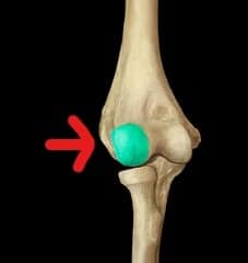
The forcible separation of the head of the radius from the structure marked in the picture below is mainly prevented by the ?
| A |
Articular capsule. |
|
| B | Annular ligament. | |
| C |
Quadrate ligament. |
|
| D |
Radial collateral ligament. |
The structure marked in the picture above represents Capitulum.
The radial notch of the ulna and the annular ligament form a ring in which the head of the radius rotates at its articulation with the capitulum of the humerus. The annular ligament narrows over the neck, cupping the head of the radius and preventing downward dislocation.
In the type of fracture shown below, which among the following is true about ulnar fracture and head of radius ?

| A |
Both ulnar fracture and head of radius is displaced posteriorly. |
|
| B |
Both ulnar fracture and head of radius is displaced anteriorly. |
|
| C |
Ulnar fractures is posteriorly and head of radius is displaced anteriorly. |
|
| D |
Ulnar fracture is anteriorly and head of radius is displaced posteriorly. |
The fracture shown in the picture above represents Monteggia fracture.
In Monteggia fracture, both ulnar fracture and head of radius is displaced anteriorly.
