
KIDNEY
| A | Rt. kidney is preferred over the left for transplantation | |
| B |
It is lower than the left kidney |
|
| C |
Rt. renal vein is shorter than the left |
|
| D |
Rt. kidney is related to the duodenum |
| A |
Rt. kidney is preferred over the left for transplantation |
|
| B |
It is lower than the left kidney |
|
| C |
Rt. renal vein is shorter than the left |
|
| D |
Rt. kidney is related to the duodenum |
Ans. is ‘a’ ie. Rt. kidney is preferred …..
It is left kidney, not Rt. that is preferred for transplantation because it has a longer renal vein which simplifies the transplantation operation.
What is column of Bertini in kidney?
| A |
Renal tumor |
|
| B |
Tongue like papillary projection |
|
| C |
Calculus |
|
| D |
None |
Ans. is ‘b’ i.e., tongue like papillary projection
The neonatal kidney achieves concentrating ability equivalent to adult’s kidney by:
| A |
One year of age |
|
| B |
Eighteen months of age |
|
| C |
Three to six months of age |
|
| D |
Just before puberty |
Answer is A (One year of age) :
“Normal adult kidney can produce urine with an osmolalitv of greater than 1000 m Osm/kg water. The healthy new born infant, subject to water deprivation for 10-14 hours can achieve an osmolality of only 500-700 m osm / kg.
This value increases gradually during the first few months and approximates to the adult value by about one year of age.”
March 2002
| A |
Dorsal region |
|
| B |
Lumbar region |
|
| C |
Thoracolumbar region |
|
| D |
Sacral region |
Ans. D i.e. Sacral region
Extent of kidney is from:
| A |
D10-L1 |
|
| B |
D11-L2 |
|
| C |
D12-L3 |
|
| D |
L1-L3 |
D12-L3
Kidney is supported by all of the following except:
| A |
Perirenal fat |
|
| B |
Renal fascia |
|
| C |
Pararenal fat |
|
| D |
Fibrous capsule |
The kidneys have the following coverings:
- Fibrous capsule: This surrounds the kidney and is closely applied to its outer surface.
- Perirenal fat: This covers the fibrous capsule.
- Renal fascia: This is a condensation of connective tissue that lies outside the perirenal fat and encloses the kidneys and suprarenal glands; it is continuous laterally with the fascia transversalis.
- Pararenal fat: This lies external to the renal fascia and is often in large quantity.
- It forms part of the retroperitoneal fat.
The perirenal fat, renal fascia, and pararenal fat support the kidneys and hold them in position on the posterior abdominal wall.
Regarding kidney, all of the following statements are true except:
| A |
They lie at the vertebral level T12 to L3 |
|
| B |
Retroperitoneal organ |
|
| C |
Left kidney is situated lower than the right |
|
| D |
Left kidney is located slightly more medial than the right |
The kidneys are reddish brown and lie behind the peritoneum high up on the posterior abdominal wall on either side of the vertebral column; they are largely under cover of the costal margin.
The right kidney lies slightly lower than the left kidney because of the large size of the right lobe of the liver.
On the medial concave border of each kidney is a vertical slit that is bounded by thick lips of renal substance and is called the hilum.
The hilum transmits, from the front backward, the renal vein, two branches of the renal artery, the ureter, and the third branch of the renal artery (VAUA). Lymph vessels and sympathetic fibers also pass through the hilum.
| A |
12th rib and lateral border of sacrospinalis |
|
| B |
11th rib and lateral border of quadratus lumborum |
|
| C |
11th rib and lateral border of sacrospinalis |
|
| D |
12th rib and lateral border of quadratus lumborum |
The angle between the lower border of the 12th rib and the outer border of the erector spinae is known as the renal angle Renal angle
- It is the angle between lateral border of Erector spinae and lower border of twelfth rib on the posterior aspect of the trunk.
- In most of the cases any abnormality (e.g., pain, tenderness, fullness, bulge) in this region is an indicative of renal origin.
| A |
Ureteric bud |
|
| B |
Mesonephros |
|
| C |
Metanephros |
|
| D |
None |
Ans. is ‘c’ i.e., Metanephros
Development of kidney
- Ureteric bud (mesonephros) arise from mesonephric duct and gives rise to collecting system of kidney (renal pelvis, major and minor calyces, collecting tubule) and ureter.
- Metanephric mesoderm (blastema or metanephors) arise from nephrogenic cord which in turn is derived from intermediate mesoderm. It gives rise to excretory unit (nephron), i.e. glomeruli, PCT, Loop of henle and DCT.
| A |
Paramesonephric duct |
|
| B |
Mullerian duct |
|
| C |
Mesonephric duct |
|
| D |
None |
Development of kidney
- Ureteric bud (mesonephros) arise from mesonephric duct and gives rise to collecting system of kidney (renal pelvis, major and minor calyces, collecting tubule) and ureter.
- Metanephric mesoderm (blastema or metanephors) arise from nephrogenic cord which in turn is derived from intermediate mesoderm. It gives rise to excretory unit (nephron), i.e. glomeruli, PCT, Loop of henle and DCT.
| A |
Metanephros |
|
| B |
Mesonephros |
|
| C |
Blastema |
|
| D |
All of the above |
All of the above
Duct of Bellini are present in ‑
| A |
Liver |
|
| B |
Spleen |
|
| C |
Kidney |
|
| D |
Lung |
Collecting duct of kidney is also called as Papillary duct or duct of Bellini.
Secretory part of kidney develops from ‑
| A |
Ureteric bud |
|
| B |
Mesonephros |
|
| C |
Blastema |
|
| D |
Paramesonephric duct |
Ans. is ‘c’ i.e., Blastema
Development of kidney
Ureteric bud (mesonephros) arise from mesonephric duct and gives rise to collecting system of kidney (renal pelvis, major and minor calyces, collecting tubule) and ureter.
Metanephric mesoderm (blastema or metanephors) arise from nephrogenic cord which in turn is derived from intermediate mesoderm. It gives rise to excretory unit (nephron), i.e. glomeruli, PCT, Loop of henle and DCT.
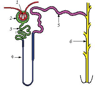
| A |
Mesonephros |
|
| B |
Metanephros |
|
| C |
Ureteric Bud |
|
| D |
Mesonephric duct |
6 segment is Collecting Tubules.
- URETERIC BUD (MESONEPHROS) arises from lower part of mesonephric duct.
- URETERIC BUD forms COLLECTING PART of kidney i.e. pelvis , Major calyces, Minor calyces, Collecting tubules, Ureter.
- METANEPHROS forms EXCRETORY UNIT of kidney i.e. Glomerulus , proximal convoluted tubule , loop of henle , distal convoluted tubule.
