
KIDNEY- STRUCTURE, RENAL FASCIA & VASCULATURE
STRUCTURE OF KIDNEY
- Parenchyma of the kidney is divided into two major structures:
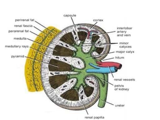
– Outer renal cortex and
– Inner renal medulla
- The medulla comprises about 10 renal pyramids. Their apices form renal papillae which indent the minor calyces.
The cortex is divided into two parts:
- Cortical arches or cortical lobules, which form caps over the bases of the pyramids.
- Renal columns, which dip in b/w the pyramid.
- Between the renal pyramids are projections of cortex called renal columns (or Bertin columns).
- Renal cortex surrounding a portion of medulla called a renal pyramid (of Malpighi).
- Each pyramid alongwith overlying cortical arch forms a lobe of the kidney.
→ RENAL SINUS: is a space that extends into kidney from the hilus.
It contains:-
– Branches of renal artery– Tributaries of renal vein– Renal pelvis: Pelvis divided into 2 to 3 major calyces, & these in their turn divide into 7-13 minor calyces.
- Each minor calyx ends in an expansion which is intended by one to three renal papillae.
→ RENAL PELVIS & CALYCES are lined by transitional epithelium.
RENAL FASCIA:
- There are 4 coverings around the kidney (from within outwards):-

1) TRUE CAPSULE (Fibrous capsule):
- It is formed by the condensation of fibrous stroma of Kidney.
2) PERINEPHRIC CAP (Adipose capsule):
- It occupies the interval b/w the fibrous capsule & renal fascia (false capsule or fascia of Gerata).
- This space (occupying perinephric fat) is called space of Gerota.
3) FALSE CAPSULE (Renal fascia or fascia of Gerota):
- It is formed by condensation of extra-peritoneal connective tissue of two layers: anterior: “fascia of Toldt” & posterior “fascia of Zuckerkendl”.
4) PARANEPHRIC FAT:
- It occupies the interval b/w the renal fascia & anterior layer of thoracolumbar fascia.
RENAL VASCULATURE:
In fetal life:
- Due to ascent of kidneys during development, the blood supply of kidney changes:-
– Initially when the kidneys are in pelvis, the renal arteries are branches of common iliac arteries.
– With progressive ascent,the arteries to kidneys are derived from different levels of aorta.
- Upto 5th week of intrauterine life, kidney is in lumbar region and renal arteries are branches of common iliac artery .
- After that, differential growth of abdominal wall causes the kidney to ascent to lumbar region. Adult position (lumbar region of abdomen) is attained by 9th week.
- Druing progressive ascent, the arteries to kidney are derived from different levels of aorta.
- After full ascent, definitive renal artery is branch of aorta at 2nd lumbar segment.
- Thus, neonatal kidney is supplied by aorta.
Arterial Supply:
- Each kidney is supplied by renal arteries, left and right, which branch from left & right phrenic artery which branch directly from the abdominal aorta.
- Posterior, apical, upper anterior, middle anterior and lower are 5 segments of vascular supply in each kidney
- Kidneys receive approximately 20% of the cardiac output.
- Renal artery→Segmental arteries→Interlobar arteries (penetrate the renal capsule and extend through the renal columns between the renal pyramids)
- Interlobar arteries supply→Arcuate arteries (run through the boundary of the cortex and the medulla) → Interlobular arteries → Afferent arterioles (supply the glomeruli).
- Branches of renal artery are end arteries.
- Right renal artery is longer and passes behind IVC.

Venous Drainage:
- Right renal vein is shorter than the left .
- Left renal vein is longer and passes in front of abdominal aorta, behind the origin of superior mesenteric artery.
- Left renal vein also receives left inferior phrenic vein, left gonadal vein, & left suprarenal (adrenal) vein.
- Each renal vein begins beneath the true capsule as stellate vein.
NERVE SUPPLY:
- Kidney is supplied by renal plexus, an offshoot of coelic plexus.
- It contains symapathetic (T10-L1) fibres which are chiefly vosomotor.
- The afferent nerves of the kidney belongs to segments T10-T12.
RENAL ANGLE
- Renal angle lies between the lower border of 12th rib and outer border of errector spinae (sacrospinalis).
Exam Important
- Right renal vein is shorter than the left
- Superior suprarenal artery originates from Inferior phrenic artery
- Initially when the kidneys are in pelvis, the renal arteries are branches of common iliac arteries
- Neonatal kidney is supplied by aorta.
- Right renal artery is longer and passes behind IVC
- There are 5 vascular segment in kidney:Posterior, apical, upper anterior, middle anterior and lower.
Don’t Forget to Solve all the previous Year Question asked on KIDNEY- STRUCTURE, RENAL FASCIA & VASCULATURE
Click Here to Start Quiz
Click Here to Start Quiz

