
Abnormalities of Placenta and Cord
Abnormalities of Placenta and Cord
PLACENTA SUCCENTURIATA:
- One (usual) or more small lobes of placenta, size of cotyledon, may be placed at varying distances from the placental margin
- In absence of communicating blood vessels, it is called placenta spuria.
- Incidence: 3%
Clinical significance:
- If the succenturiate lobe is retained, following birth of the placenta, it may lead to:
- Postpartum haemorrhage
- Subinvolution
- Uterine sepsis
- Polyp formation
Treatment:
- Missing lobe :exploration of the uterus and removal of the lobe under general anaesthesia

PLACENTA EXTRACHORIALIS
TYPES:
1.Circumvallate placenta:
- Development:due to smaller chorionic plate than the basal plate resulting from Recurrent marginal hemorrhage
- Morphology:
- Fetal surface is divided into a central depressed zone surrounded by a thickened white ring (complete)
- Vessels radiate from cord insertion as far as the ring and then disappear from view.
- The peripheral zone is thicker and the edge is elevated and rounded

2.Placenta marginata:
- Thin fibrous ring present at margin of chorionic plate where the fetal vessels appear to terminate.

PLACENTA MEMBRANECEA:
- Placenta is unduly large and thin
- Develops from chorion frondosum & chorion laeve
- Clinical significance:
- Enroachment of some part over lower segment
- Imperfect separation in third stage
- Chance of retained placenta
- Manual removal difficult.

- Abnormalities in which trophoblastic tissues invade the myometrium to varying depths.
- Much more likely with placenta previa or with implantation over prior uterine incision or perforation.

CORD ABNORMALITIES:
SHORT CORD:
- Less than 20cm
- Due to entanglement of cord round any fetal part.
- Exceptionally: cord may be absent and placenta may be attached to liver as in exomphalus.
- Clinical significance:
- Prevent descent of presenting part
- Separation of normally situated placenta
- Favour malpresentation
- Acute inversion
- Fetal growth restriction
- Intrapartum distress
- Failure of external version
- Two fold risk of fetal death

LONG CORD:
- Clinical Significance
- Cord prolapse
- Cord entanglement round the neck or the body
- True knot
- False knots
SINGLE UMBILICAL ARTERY:
Incidence :
- 0.63 %in liveborns
- 1.92 % in perinatal deaths
- 3 %in twins.
- Common in twins and in babies born of diabetic mothers or in polyhydraminos.
Frequently association:
- Congenital malformation of fetus (10-20%).
- Renal and genital anomalies
- Trisomy 18
Increased chance of :
- Abortion
- Prematurity
- FGR
- Perinatal mortality
Diagnosis:
- Sonographic screening

BATTLEDORE PLACENTA:
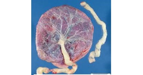
- Cord attached to margin of the placenta
- If associated with low implantation of the placenta, chances of cord compression in vaginal delivery leading to fetal anoxia or even death.

VELAMENTOUS PLACENTA:
- Umbilical vessels spread within the membranes distance from the placental margin
- They reach surrounded only by a fold of amnion.
- Risk of Fetal exsanguinations before labor
- Common in placenta previa and multifetal gestations

VASA PRAEVIA:
- Leash of blood vessels happen to traverse through the membranes overlying the internal os & can be ruptured easily.
- Incidence is 1:1500
- Associated with low lying placenta
MANAGEMENT:
- Fetal bleeding: urgent delivery (vaginally or CS )
- The newborn’s haemoglobin is estimated and if necessary, blood transfusion be carried out.
- If the baby is dead, vaginal delivery is awaited.
BIPARTITE PLACENTA:
- Two complete and separate parts are present, each with cord leaving it.
- The bipartite cord joins short distance from the two parts of the placenta
- Tripartite placenta is similar but with three distinct parts

CORD ABNORMALITIES CAPABLE OF IMPENDING BLOOD FLOW:
KNOTS:
False Knots
- Knobs protruding from cord surface and are focal redundancies of a vessel or Wharton jelly, with no clinical significance.
True Knots
- Active fetal movements create cord knotting
- Stillbirth risk

- Cord frequently becomes coiled around portions of fetus.
- Those looped around neck are termed nuchal cord(20 – 34% of deliveries)
- Two loops: 34%
- Three loops: 0.2 – 0.5%
FUNIC PRESENTATION:
- Cord prolapse or fetal heart rate abnormalities is an associated labor finding.
- Diagnosis:Sonography and color flow Doppler.
- Treatment:During labor, cesarean delivery
UMBILICAL CORD STRICTURE:
- Focal narrowing of cord diameter
- Develops in area of fetal umbilical insertion
- Absence of Wharton jelly and stenosis or obliteration of cord vessels at the narrow segment
- Most fetuses are stillborn
Exam Important
- The findings of a single umbilical artery on examination of the umbilical cord after delivery is An indicator of considerably increased incidence of major malformation of the fetus
- Velamentous insertion of the cord is associated with an increased risk for Fetal exsanguinations before labor
- Placenta in which vessels seperate before reaching margin is Velamentous placenta
- Fetal blood loss in abnormal cord insertion is seen in Vasa previa
- Single umbilical artery is associated with diabetes in mother.
- Failure of development of one artery in later months is implicated single umbilical artery case
- Vasa previa is Associated with low lying placenta
- In vasa previa Incidence is 1:1500
- In vasa previa Caesarian section is indicated
- Single umbilical artery is associated with Polyhydramnios, Fetal growth retardation & Increased incidence of fetal malformation
- Unilateral renal agenesis is associated with Single umbilical artery
Don’t Forget to Solve all the previous Year Question asked on Abnormalities of Placenta and Cord

