

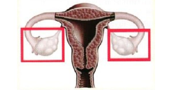
Anatomy Of Ovaries
DEVELOPMENT:
- Formation of primary ovary in female foetus takes place by 10th week
- Coelomic epithelium on medial side of the mesonephros becomes thickened to form genital ridge, the site where ovary develops.
- Genital ridge is covered by germinal epithelium (previous coelomic epithelium). From these germinal epithelium, cords of cells (sex cords or medullary cords) proliferate and grow into the underlying mesoderm.
- Primordial germ cells which are developed from endodermal cells of hindgut (part of yolk sac), migrate to region of developing ovary (genital ridge area) and give rise to oocytes.
- The sex cords become broken up into small masses. The cells of each mass surround one oocyte to form primordial follicle.
- At birth each ovary contains about 2 million primary follicles.
RELATION:
- POSTERIOR:Attached to the back of the broad ligament by mesovarium.
- LATERAL: Fossa below the bifurcation of the common iliac artery and the ureter
- MEDIALLY:Fimbria of the fallopian tube
BORDERS:
- Anterior border: (Mesovarian border) is straight and related to fallopian tube and obliterated umblical artery.Attached to posterior surface of broad ligament of uterus by mesovarium and forms the hilus of ovary.
- Posterior border :Free border is convex and is related to fallopian tube and ureter.
ATTACHMENTS:
- Cornu of the uterus by the ovarian ligament
- The suspensory ligament attaches the ovary to the pelvic wall and carries within its folds the main portion of the ovarian vessels and nerves.

COMPONENTS:
The ovary has 3 components;
- Surface: The surface layer of the ovary is formed by simple cuboidal epithelium, known as germinal epithelium.
- Cortex: The cortex (outer part) of the ovary is largely comprised of a connective tissue stroma.
- It supports thousands of follicles.
- Each primordial follicle contains an oocyte surrounded by a single layer of follicular cells.
Medulla:
- The medulla (inner part) is composed of supporting stroma and contains a rich neurovascular network which enters the hilum of ovary from the mesovarium.
- Cells found in the interstitium of the ovary that resemble steroid secreting cells and may secrete androgens in the human ovary are Ovarian hilar cells

BLOOD AND NERVE SUPPLIES:
- The ovaries receive blood from paired ovarian arteries, which arise directly from the abdominal aorta, below the renal artery.
- Venous drainage is achieved by a pair of ovarian veins. The left ovarian vein drains into the left renal vein, and the right ovarian vein drains directly into the inferior vena cava.
- Lymph from the ovaries drains into the Preaortic & para-aortic nodes.
- The ovaries receive sympathetic and parasympathetic nerve fibres from the ovarian and uterine (pelvic) plexuses, respectively
- The nerve supply of the ovaries arises from T10.

OVARIAN FOSSA:
- Each ovary lies in ovarian fossa on lateral pelvic wall which is bounded 😕
- Anteriorly: Obliterated umbilical artery
- Posteriorly: Ureter and internal iliac artery
Exam Question
- Ovary develop from Genital ridge
- The main portion of the ovarian artery, vein, and nerves are carried to and from the ovary by way of the Suspensory ligament
- The nerve supply of the ovaries arises from T10.
- Cells found in the interstitium of the ovary that resemble steroid secreting cells and may secrete androgens in the human ovary are Ovarian hilar cells
- Formation of primary ovary in female foetus takes place by 10th week
- Germ cells in the ovary develop from Yolk sac
- Lymphatic drainage of ovary is through Preaortic and para-aortic lymph nodes
- At birth each ovary contains about 2 million primary follicles.
- Sex cords are derived from coelomic epithelium
- Ovarian fossa is formed by Obliterated umbilical artery, Internal iliac artery & Ureter
- Posterior border of ovary is Free of ligament attachment
- Primordial Germ cell of ovary are developed from Endoderm
Don’t Forget to Solve all the previous Year Question asked on Anatomy Of Ovaries
