


CVS MONITORING IN ANESTHESIA
Introduction:
Methods:
- Non-invasive.
- Invasive
Non-invasive methods:
ECG:
- Excellent monitor to detect arrhythmia, ischemia/cardiac arrest.
- Arrhythmia detection – By lead II.
- Ischemia detection – By lead V2.
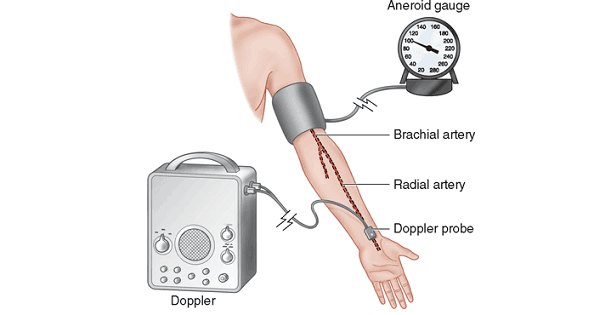
Non-invasive B.P. (NIBP):
- NIBP instruments measure BP automatically at regular interval.
- Important factor – Cuff size.
- Should cover 2/3rd of arm length.
- Too large cuff will under-read actual value.
- Too small cuff will over-read actual value.
Transesophageal echocardiography:
2-dimensional TEE:
- Rapid & more sensitive indicator of myocardial ischemia than ECG.
- Detects myocardial ischemia & air embolism in peri-operative period.
- Detects regional wall motion abnormality.
- Decreased systolic wall thickening – More reliable index for ischemia than endocardial wall motion abnormality.
Invasive methods:
1. Invasive blood pressure (IBP):
- Recorded by transducer inside artery.
- Radial artery – Most common artery.
- Dorsal pedis artery – In infants.
Intra-arterial cannulation:
- Arterial blood sampling (ABG measurement).
- Intra-arterial BP measurement.
- Drug injection.
- Blood transfusion.
2. Central venous pressure monitoring (CVP):
- Most commonly in right internal jugular vein.
- Measured in subclavian, basilic & femoral veins.
- Normal CVP = 3-10 cm of H20 (2-8 mm Hg).
Increased CVP:
- CHF, IPPV with PEEP & Valsalva maneuver.
Decreased CVP:
- Venodilators (nitroprusside).
Indications:
- Estimation of intravascular volume – Guide to fluid therapy mainly blood/plasma transfusion.
Complication:
- Air embolism, hemothorax, sepsis & trauma to carotid artery (cause pseudoaneurysm).
3. Pulmonary artery catheterization:
- Catheter insertion into pulmonary artery.
- Catheter used – Swan-Ganz catheter.
Uses:
- Direct simultaneous measurement of pressure in right atrium, right ventricle & pulmonary artery.
- Cardiac output.
- Oxygen saturation of mixed venous blood – Pulmonary artery (best site).
Exam Important
- Non-invasive B.P. (NIBP) important factor is cuff size & should cover 2/3rd of arm length, too large cuff will under-read actual value & too small cuff will over-read actual value.
- Transesophageal echocardiography (2-dimensional TEE) is rapid & more sensitive indicator of myocardial ischemia than ECG.
- 2-dimensional TEE is detecting myocardial ischemia & air embolism in peri-operative period & detects regional wall motion abnormality.
- 2-dimensional TEE is decreased systolic wall thickening is more reliable index for ischemia than endocardial wall motion abnormality.
- Invasive blood pressure (IBP) is usually recorded in most common artery is radial artery.
- Central venous pressure monitoring (CVP) is normal CVP = 3-10 cm of H20 (2-8 mm Hg).
- CHF, IPPV with PEEP & Valsalva maneuver are conditions increasing CVP.
- Venodilators (nitroprusside) is decreasing CVP.
- Estimation of intravascular volume is a guide to fluid therapy mainly blood/plasma transfusion.
- Complications of CVP are air embolism, hemothorax, sepsis & trauma to carotid artery (cause pseudoaneurysm).
- Pulmonary artery catheterization is done by Swan-Ganz catheter.
- Pulmonary artery catheterization measures direct simultaneous measurement of pressure in right atrium, right ventricle & pulmonary artery.
- Pulmonary artery catheterization is used for measurement of cardiac output & measuring oxygen saturation of mixed venous blood.
Don’t Forget to Solve all the previous Year Question asked on CVS MONITORING IN ANESTHESIA

