
DEVELOPMENT OF HEART
Development of Heart
- Develops from splanchnic mesoderm forming the cardiogenic area.
- Primordial heart develops as two endothelial heart tubes which fuse to form a single tubular heart.
- Heart tube (tubular heart) is formed at the end of 3rd week.
- Endothelial heart tube is surrounded by a layer of mesoderm known as myo-epicardial mantle.
- Myo-epicardial mantle gives rise to myocardium & visceral pericardium (epicardium).
- Myo- epicardium secretes a thick layer of extracellular matrix (hyaluronic acid) c/d cardiac jelly that separates it from tubular heart.
Developing heart consists of 3 layers:
- Endocardium
- Myocardium
- Epicardium (serous pericardium)
Tubular heart has following components, cranio-caudally :
1. Bulbus cordis:
Cranial most part & subdivided into:
- Truncus arteriosus (distal part)
- Conus cordis (mid portion)
- Proximal part
2. Primitive ventricle:
- Along with conus cordis it forms the right & left ventricles.
3. Primitive atrium:
- Later form right & left atria.
4. Sinus venosus
- Caudal most part of tubular heart.
- As its lower end presents right & left horns.
- Each horn receives blood from following three veins:
- Vitelline vein from yolk sac. Right vitelline vein forms terminal part of inferior vena cava.
- Umbilical vein from placenta
- Common cardinal vein from body wall. Right common cardinal vein forms superior vena cava.
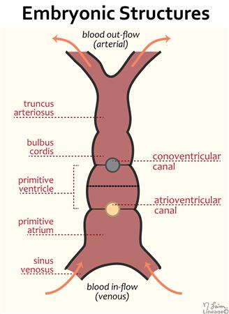
→ Truncus arteriosus is the arterial end & sinus venosus is venous end of heart tube (tubular heart).

FORMATION OF ATRIA
- Formation of atria involves following processes:
1. Septation of atrioventricular canal (AV canal)
- AV canal is the passage through which primitive atrium is connected to primitive ventricle.
- AV canal is divided into right & left canal by endocardial cushions (atrioventricular cushions or AV cushions).
- The fused cushions form septum intermedium.
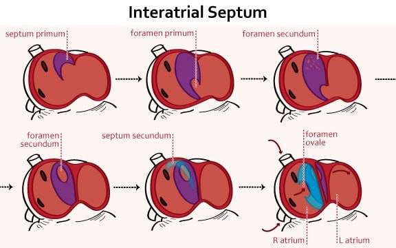
2. Septation of primitive atrium:
- The primitive atrium divided into left & right atria by interatrial septum.
- Formed by septum primum & septum secundum.
- Septum secundum grows caudally from the roof of atrium & overlaps the foramen secundum.
3. Incorporation of sinus venosus into right atrium:
- Right horn of sinus venosus is incorporated to form posterior smooth part of right atrium.
- Left horn of sinus venosus forms the coronary sinus.
4. Incorporation of pulmonary veins into left atrium
- Right & left tributaries & their first bifurcations, are incorporated to form posterior smooth part of left atrium.
FORMATION OF VENTRICLES
1. Right ventricle
Develops from:
- Rough part: from proximal part of bulbar cordis & right half of primitive ventricle.
- Smooth part (infundibulum): from caudal part of bulbus cordis (conus cordis).
2. Left ventricle
Develops from:
- Rough part: from left half of primitive ventricle.
- Smooth part (vestibule): from caudal part of bulbus cordis (conus cordis).
- Intraventricular septum separates right ventricle from left ventricles.
Formation of intraventricular septum involves 3 processes:
- Formation of proximal bulbar septum, which separates conus cordis part.
- Formation of muscular part of interventricular septum.
- Formation of membranous part of interventricular septum.

→ Heart is completely developed at 10th week & very much resembles that of a newborn.
Exam Important
- Tubular heart is formed at the end of 3rd week.
- Septum secundum arises from Primitive atrium.
- The cardiac jelly formed around the heart tube during early development, contributes to the formation of Endocardial cushion valves.
- Septum secundum grows caudally from the roof of atrium.
- Fossa ovalis and anulus ovalis lie on the atrial septum, which separates the right atrium from the left atrium.
- Heart is completely developed at 10th week of intrauterine life.
