
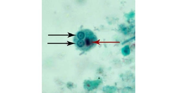
Entamoeba Histolytica: Clinical Manifestations
Mode of Transmission:
- Faecal-oral route
- Oral-rectal contact
- Transmission through vectors ( flies, cockroaches and rodents)
- Man main reservoir
Incubation period:
- Varies a great deal depending upon the host’s resistance to the parasite.
- Generally, it is four to five days
Primary or intestinal lesions:
- Primary infection of this parasite is limited entirely to the large intestine.
1. Amoebic dysentery:
- Parasite presence and activity of in wall of large intestine
Amebiasis most frequently involves :
- the cecum and ascending colon
- followed in order by the sigmoid, rectum, and appendix.
- Causes amoebic dysentery characterised by frequent passage of stool and griping pam.
- The contain mucus and occasionally blood.
2. Amoebic ulcers:
- Multiple ulcers in the caecum, ascending colon and rectum.
- In advanced cases, haemorrhage due to erosion of blood vessels.
- The ulcerative lesions develop undermining of margins of the ulcer due to lytic action of the trophozoites.
- Ulcer has a flask shaped with a narrow neck and broad base.
3. In acute case :
- Perforation and gangrene of intestinal
- Pericolic abscesses & peritonitis
Metastatic or secondary lessions:
- Enter into the blood circulation
- Reaches the different parts of the body
- Cause extra intestinal or metastatic lesion.
- It is also known as “tissue amoebiasis”.
1.Amoebic liver abscess :
- May be single large abscess or multiple, and may involve both lobes.
- In the tropics 2-10% of the individuals infected with E. histolytica suffer from hepatic complications
Common in :
- Most common in right lobe liver/right posterior-superior region
- Portal vein is in direct continuation with the right tributary.
- Pus in amoebic liver abscess is not due to suppuration, but is a mixture of sloughed liver tissue and blood
- Pus is chocolate coloured, classically called anchovy-sauce
- Abscess wall good for culture
Complications
- Pleuropulmonary infection is the most common complication.
- Cardiac involvement results following the rupture of an abscess involving the left lobe of the liver.
- Associated with very high mortality.
- Intraperitoneal rupture occurs in 2-7% of patients.
- Left lobe abscesses are more likely to progress to rupture because of their later clinical presentation
2. Pulmonary amoebiasis:
- Appearance of single or multiple abscesses in one or both lungs.
- An amoebic lung abscess is almost always secondary to spread from the liver.
3. Cerebral amoebiasis:
- Appearance of small absess in one of the cerebral hemisphere.
4. Splenic amoebiasis:
- Rare
- Cause abscess.
5. Cutaneous amoebiasis:
- Skin lesions
- Skin is involved directly over the region adjoining a visceral lesion
- Extensive necrosis and sloughing of the skin and subcutaneous tissues are caused by the trophozoites of E. histolytica
- Granulomatous ulceration and granulomatous mass may occur in the peri-anal region.
6. Urinogenital tract Infection:
- Rare
- Cause amoebic ulcer of the penis and vaginitis.
Exam Question
- The main reservoir for entamoeba histolytica is man
Mode of Transmission:
- Faecal-oral route
- Oral-rectal contact
- Transmission through vectors ( flies, cockroaches and rodents)
- Amebiasis most frequently involves the cecum and ascending colon, followed in order by the sigmoid, rectum, and appendix.
Cutaneous amoebiasis
- Direct infection from liver
- Granulomatous ulceration and granulomatous mass may occur in the peri-anal region.
Characteristic feature of amoebic ulcer
- Flask shaped
- Undermined edge
Amoebic liver abscess :
- In the tropics 2-10% of the individuals infected with E. histolytica suffer from hepatic complications
- Liver abscess is the most common extra-intestinal manifestation of amoebiasis.
Common in :
- Most common in right lobe liver/right posterior-superior region
- Portal vein is in direct continuation with the right tributary.
- Pus in amoebic liver abscess is not due to suppuration, but is a mixture of sloughed liver tissue and blood
- Pus is chocolate coloured, classically called anchovy-sauce
- Abscess wall good for culture
- Pleuropulmonary infection is the most common complication.
- Most common cause of amoebic lung abscess is direct spread from liver
- Amoebic liver abscess ruptures most commonly into pleural cavity
Don’t Forget to Solve all the previous Year Question asked on Entamoeba Histolytica: Clinical Manifestations
