
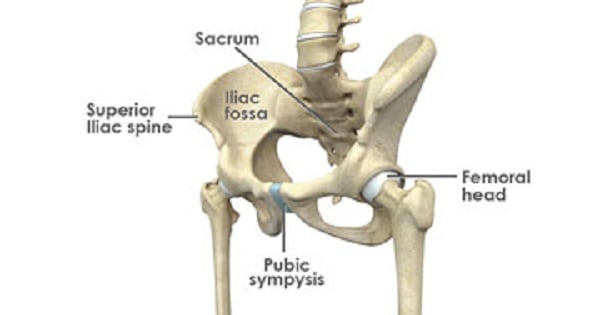
Hip joint
HIP JOINT
- Body’s second largest weight-bearing joint (after the knee).
- Multiaxial “ball and socket” joint at the juncture of the leg and pelvis.
- The rounded head of the femur (thighbone) forms the ball, which fits into the acetabulum (a cup-shaped socket in the pelvis).
- Ligaments connect the ball to the socket and usually provide tremendous stability to the joint.
- The hip joint is normally very sturdy because of the fit between the femoral head and acetabulum as well as strong ligaments and muscles at the joint.
- All of the various components of the hip mechanism assist in the mobility of the joint.
BONES OF HIP JOINT:
- The femur is the upper leg bone or thigh. It is the largest bone in the body.
- At the top of the femur is a rounded protrusion which articulates with the pelvis. This portion is referred to as the head of the femur, or femoral head.
- There are two other protrusions near the top of the femur, known as the greater and lesser trochanters. The muscles involved in hip motion are attached to the joint at these trochanters.
- The acetabulum is a concave area in the pelvis, into which the femoral head fits.
- The pelvis is a girdle of bones, connected at the front by cartilage pad, called the pubis, and at the back by the lowest four fused vertebrae (the sacrum).
- The sacro-iliac joints are located where the sacrum meets the pelvis.
- The bone surfaces of the femoral head and acetabulum have a smooth durable layer of articular cartilage that cushions the ends of the bones and allows for smooth movement.
HIP JOINT CAPSULE OR SOCKET:
- The joint capsule is a thick ligamentous structure surrounding the entire joint.
- Inside the capsule, the surfaces of the hip joint are covered by a thin tissue called the synovial membrane.
- This membrane nourishes and lubricates the joint.
- Stability of the hip joint is directly related to its muscles and ligaments.
The most notable ligaments in the hip joint are:
LIGAMENTS:
A. Intracapsular ligaments:-
- These ligaments are inside the capsule. These are:
- Acetabular labrum (labrum acetabular): Deepens the acetabulum & is deficient inferiorly.
- Transverse acetabular ligament: Bridges the acetabualr notch on the inferior margin of acetabulum.
- Ligament of head of femur (logamentum teres or round ligament): Connects transverse acetabular ligament to hte fovea of femoral head.
B. Fibrous capsule:
- Attached medially to hip bone around the margin of acetabulum.
- Laterally it is attached to intertrochanteric line on the anterior aspect & 2 cm medial (proximal) to intertrochanteric crest on posterior aspect of femoral neck.
- Hence entire anterior surface & medial half of posterior surface of femoral neck are intracapsular.
C. Extracapsular ligaments:
- These ligaments are outside the capsule. These are:
1. Iliofemoral ligament (Y-ligament of Bigelow):
- Strongest ligament of the body.
- The stem of ‘Y’ is attached above to anterior superior iliac spine.
- Strongly resist hyperextension of the hip joint, & thereby prevents the trunk falling backwards.
- Its medial band limits abduction & lateral band limits adduction.
2. Pubofemoral ligament:
- Covers anteromedial surface of capsule.
3. Ischiofemoral ligament:
- Covers the capsule posteriorly & prevents hyperextension of hip joint to asssist iliofemoral ligament.
LABRUM:
- The labrum is a circular layer of cartilage which surrounds the outer part of the acetabulum effectively making the socket deeper to provide more stability for the joint.
MUSCLES GROUP:
- The various muscles which attach to or cover the hip joint generate the hip’s movement.
1. Gluteals:
- The gluteals are the muscles in your buttocks.
- The gluteals (gluteus maximus, gluteus minimus and gluteus medius) are the three muscles attached to back of the pelvis and insert into the greater trochanter of the femur.
- Gluteus medius & minimus both causes medial rotation & abduction.
2. Quadriceps:
- The four quadricep muscles (vastus lateralis, medialis, intermedius and rectus femoris) are located at the front of the femur.
- All four attach to the top of the tibia.
- The rectus femoris originates at the front of the ilium.
- The three other quads attach around the greater trochanter of the femur and just below it.
3. Iliopsoas:
- This is the primary hip flexor muscle.
- The three parts of the iliopsoas attach the lower part of the spine and pelvis, then cross the joint and insert into the lesser trochanter of the femur.
4. Hamstrings:
- The three muscles at the back of the thigh are called the hamstrings.
- All three attach to the lowest part of the pelvis.
5. Groin muscles:
- The groin or adductor muscles attach to the pubis and run down the inside of the thigh.
NERVE SUPPLY
- Hip joint is supplied by:
- Femoral nerve
- Anterior division of obturator nerve
- The nerve to quadratus femoris
- The superior gluteal nerve
Exam Question
- Iliopsoas is the primary hip flexor muscle.
- Iliofemoral ligament, Ischiofemoral ligament, Pubofemoral ligament keeps the hip from hyper-extension.
- Bigelow’s ligament is a ligament of hip joint
- Medial rotation and Abduction is caused by Gluteus medius and Gluteus minimus
Don’t Forget to Solve all the previous Year Question asked on Hip joint
