

LEGG-CALVE-PERTHES’ DISEASE (Coxa plana, Pseudocoxalgia)
LEGG-CALVE-PERTHES’ DISEASE
The disease also known by different names such as:
- osteochondritis deformans juvenilis
- pseudocoxalgia
- coxa plana
- Occur in children 5–15 years.
- Characterized by varying degrees of idiopathic avascular necrosis of the upper femoral epiphysis.
- Followed by spontaneous revascularization over a period of time.
- During this period, the head is exposed to a risk of deformation.

Predisposing Factors
- Genetic
- Environmental factors
- Trauma
- Transient synovitis
- Passive smoking
- Abnormal venous drainage
- Arterial block and infarction
- Increased viscosity of the blood
Pathology
- 1st stage—Stage of synovitis
- 2nd stage—Stage of avascular necrosis
- 3rd stage—Stage of (fragmentation) revascularization
- 4th stage—Residual/healed stage
- Radiological Features
Based on Elizabethtown Staging:
- Stage I (Initial/Avascular necrosis): Waldenstrom’s sign
- Stage II (Fragmentation): Shrinkage and fragmentation of the epiphysis
- Stage III (Regeneration): Fragmentation along with new bone formation
- Stage IV (Healed): Complete re-ossification
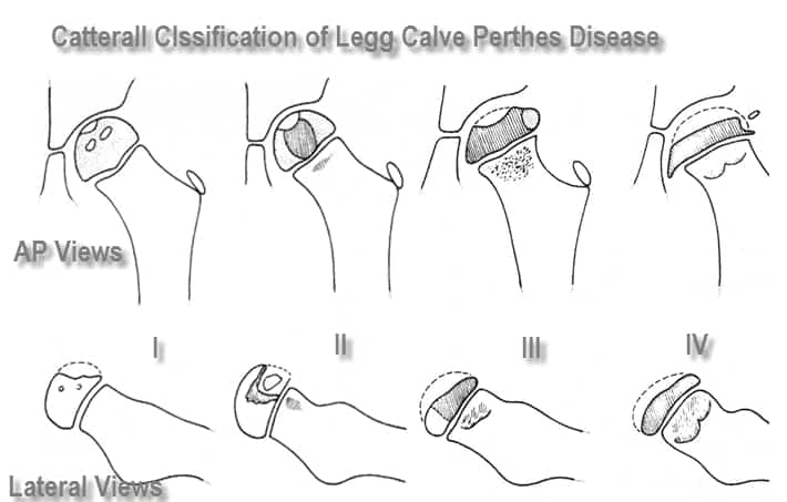
Based on Catterall Grouping:
- The disease is grouped into 4 groups depending on:
- Extent of epiphyseal involvement.
- Presence of sequestrum.
- Presence of collapse.‘Head within the Head’
Metaphyseal abnormality
Clinical Features
- Onset is insidious.
- Limp with or without dull pain.
- Pain in the hip region.
- Pain is exaggerated by activity, relieved by rest.
- Classical limitation of abduction and internal rotation.
- Trendelenburg gait may be present.
Head at Risk Signs
- Clinical “Head at Risk” Signs
- Older child
- Heavy child
- Progressive loss of movement
- Adduction contracture
- Flexion with adduction deformity.
Radiological “Head at Risk” Signs
1. Gage sign
- ‘V’ shaped radiolucent defect on the lateral side of the epiphysis.
2. Calcification lateral to the epiphysis.
- Occurs due to thickened and extruded epiphysis.
- Indicates epiphyseal enlargement.
3. Lateral subluxation.
4. Horizontal growth plate.
5. Diffuse metaphyseal lesion.


Investigations
1. X-ray:
- Avascular changes as per stage of the disease.
2. Bone scan:
- Can diagnose the avascularity much before the radiological changes develop.
- Also useful in differentiating septic from aseptic inflammation.
3. MRI:
- Delineates the changes in the cartilaginous epiphysis and the joint much better than X-ray.
- Also useful to assess vascularity of the epiphysis.
4. Arthrography:
- Useful in the assessment of joint congruity
- Helps in planning the containment procedure.
Exam Important
- Perthe’s disease is Osteochondritis of the epiphysis of the Femoral head.
- Abduction & internal rotation movements is restricted in Perthe’s disease.
- Investigation of choice for evaluation of suspected Perthes’ disease is Magnetic Resonance Imaging (MRI).
- Radiological sign in case of Perthe’s disease is Organized calcification.
Don’t Forget to Solve all the previous Year Question asked on LEGG-CALVE-PERTHES’ DISEASE (Coxa plana, Pseudocoxalgia)

