
Lymphoma
LYMPHOMA
- Most common site of extranodal lymphoma — Stomach
- Most common type — extranodal marginal zone B-cell lymphomas
- Otherwise called Mucosa associated lymphoid tissue or MALTomas
- Usually arise at the site of chronic inflammation
- Most common cause of pro-lymphomatous inflammation — H pylori
- H pylori eradication results in durable remissions and low rates of recurrence
- Translocations: t(11; 18) — most common; others — t(1;14), t(14;18)
Diagnostic histologic features
- Lympho-epithelial lesions
- Monocytoid change
- Burkitt’s lymphoma is a tumor of mature B cells that express surface IgM, CD19, CD20, CD10, and BCL6, a phenotype consistent with a germinal center B-cell origin.
- Unlike other tumors of germinal center origin, Burkitt lymphoma almost always fails to express the antiapoptotic protein BCL2
- Burkitt’s lymphoma is a B-cell lymphoma
- As this a tumor of mature B-cells it expresses IgM, CD 19, CD20, CD 10 and BCL 6 a phenotype consistent with a B-cell origin.
- However, unlike other tumors of germinal center origin, Burkitt lymphoma almost always fails to express the antiapoptotic protein BCL-2.
- All forms of Burkitt lymphoma are associated with translocations of c-Myc gene on chromosome 8.
- The usual translocation is t (8 : 14).
- Other less common translocations are t (2 : 8) and t (8 : 22).
- Most of the patients in United States with Burkitt’s lymphoma present with peripheral lymphadenopathy or an intro abdominal mass.
- The disease is typically rapidly progressive and has a propensity to metastasize to CNS.
- Chemotherapy is the treatment of choice in Burkitt’s Lymphoma.
- Burkitt’s Lymphoma was one of the first cancers shown to be curable by chemotherapy.
- Molecular pathogenesis of Burkitt’s lymphoma
- All forms of Burkitt’s lymphoma are associated with translocation of the c-MYC gene an chromosome 8.
- The translocation partener is usually IgH locus on chromosome 14, i.e., t (8; 14); But may also be Igk on chromosome 2, i.e., t (2; 8) or IgX on chromosome 22, i.e., t (8; 22).
Mantle cell lymphoma
- Mantle cell lymphoma is a type of non-hodgkin lymphoma characterized by presence of tumor cells which closely resemble the normal mantle zone of B-cells that surround germinal centers.
- Immunophenotype of mantle cell lymphoma
- Mantle cell lymphoma is neoplasm of B cells.
- Therefore it expresses B cell marker :
- CD19,CD20
- Surface immunoglobulin heavy chain (IgM and IgD).
- Either /c or X light chain.
- As the tumor cells are derived from Mantle zone, they are positive for B cell marker of mantle zone i.e., CD-5.
- Mantle cell lymphoma is CD23 negative, this feature distinguish it from chronic lymphocytic leukemia (CLL) which is positive for both CD5 and CD23.
- The other characteristic marker of mantle cell lymphoma is cycline DI.
Cytogenetic abnormalities.
- Mantle cell lymphoma is associated with an 11 : 14 translocation involving the IgH locus on chromosome 14 and the cyclin DI locus on chromosome 11.
- This leads to increased expression of cyclin D 1, which promotes GI to S phase progression during the cell cycle.
- 65 years old man with splenomegaly, lymphodenopathy CD-23 negative and CD-5 positive B-cell suggest the diagnosis of mantle cell lymphoma.
Clinical features of mantle cell lymphoma
- It is usually present in fifth to sixth decade with male preponderance.
- The most common presentation is painless lymphodenopathy.
- Splenomegaly may occur.
- Occasionally, multifocal mucosal involvement of the small bowel and colon produces lymphomatoid polyposis→ of all forms of NHL, mantle cell lymphoms is most likely to spread in this fashion.
- Prognosis of hodgkin lymphoma mainly depends on the histological type and staging.
Histological type
| Best prognosis | Lymphocytic predominent |
| Worst prognosis | Lymphocytic depletion type |
|
Best Prognosis |
Lymphocytic predominance type |
|
Worst prognosis |
Lymphocytic depletion type |
|
Most common HL |
Nodular sclerosis type |
| Most common type HL in India | Mixed cellularity type |
| Least common type HL | Lymphocytic depletion type |
Post transplant lymphoma is most commonly associated with Epstein-Barr virus
| Oncogenic DNA virus | Oncogenic RNA virus |
| Human papilloma virusQ | Hepatitis C virusQ |
| Epstein-Barr virusQ | Human T-cell leukemia virus type-1Q |
| Hepatitis B virusQ | Helicobacter pyloriQ |
Epstein-Barr Virus
- Epstein-Barr virus infection may lead to the following clinical conditions:
- Infectious mononucleosis
- EBV-associated malignancies:
- Burkitt’s lymphomaQ
- Lymphomas in immunodeficient persons such as AIDS patients and transplant recipientsQ
- Nasopharyngeal carcinoma in persons of Chinese origin
Post-Transplant Lymphoproliferative Disorder (PTLD)
- PTLD is associated with replication of EBV in B cells induced by enhanced immunosuppression, primarily observed
- in patients who have received more than one course of polyclonal antilymphocyte globulin (ALG) or monoclonal
Clinical Features:
- Clinical presentation of PTLD includes fever, malaise and lymphadenopathyQ
Diagnosis:
- The diagnosis is made by tissue biopsyQ
Treatment:
- Polyclonal PTLD: Discontinuation of immunosuppression and antiviral therapyQ
- Monoclonal PTLD: Radiation, chemotherapy and occasionally surgical resection. Antibody against CD20Q
- represents a novel approach in treating monoclonal PTLD with favorable outcome.Q
(LCA or CD45):
- “LCA or CD45 is expressed brightly in normal lymphocytes as well as in most of the lymphoproliferative disorder. It is also variably expressed in other leukocytes. It is absent in erythroid cells.” -Iochims Lymph Node Pathology 4/43
- S-100: S-100 is a marker that comes positive in chondrocytes, schwann cells, fat cells, oligodendrocytes and other neural crest origin cells and tumors.
- HMB-45: HMB-45 is a activated melanocytic marker expressed in melanoma cells. Cytokeratin: Cytokeratin is expressed in normal and malignant epithelial cells.
| Choice |
Cancers associated |
| Sjogren syndrome | NHL mainly MALT-oma involving salivary glands>stomach |
| Ataxia telengectasia | Elevated incidence of cancers, approximately 100-fold in comparison to the general population. In children, more than 85% of neoplasm cases are acute lymphocytic leukemia or lymphoma. In adults with ataxia-telangiectaisa, solid tumors are more frequent |
| Lynch-II syndrome | Gastrointestinal cancer associated with endometrial/ovarian carcinoma. Early onset brain tumor and lymphoma also seen in children
Lymphoma in Sjogren’s syndrome The development ofLymphomas in patients with Sjogren syndrome is suggested by : –
|
- he stomach is the most frequent extranodal site for lymphomas. Microscopically, the vast majority of gastric lymphoid tumors are non-Hodgkins lymphomas of B cell origin, Hodgkins ds involving the stomach is extremely uncommon.”
-
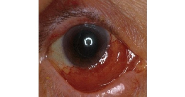
Most patients notice the reddish discoloration of the surface of the eyeball (conjunctiva).
As the tumor enlarges, patients seek medical attention.
Conjunctival lymphomas can become large enough to displace the eyeball, and restrict eye movement. Eye movement restriction can cause diplopia (double-vision).
If the tumor extends behind the eyeball, it can be pushed forward (proptosis). Rarely, and if large enough, orbital lymphoma can press on the optic nerve and cause loss of vision.
If discovered early, prompt treatment offers the best chance for recovery of vision.
-
n 1964, researchers at the National Cancer Institute developed the first combination chemotherapy that cured a number of patients who had relapsed following a standard radiation therapy regimen.
This drug combination was called MOPP and was, for a long time, the standard treatment for Hodgkin’s disease:
- Mustargen (mechlorethamine, nitrogen mustard)
- Oncovin (Vincristine, VCR)
- Procarbazine (Matulane)
- Prednisone (Deltasone, Orasone)
- MOPP has been mostly replaced by another combination chemotherapy called ABVD (Adriamycin, Bleomycin, Vinblastine, Dacarbazine), which is now the standard chemotherapy regimen for Hodgkin’s disease.
- However, MOPP may be used if there are lung or heart conditions present or allergies to any of the medications in the ABVD combination. Additionally, 30% to 40% of people will relapse after treatment with ABVD, which will require ‘salvage’ treatment with MOPP.
- VAD combination chemotherapy is used for multiple myeloma and CHOP combination chemotherapy is used for B cell chronic lymphoid leukemia
- Reed-Sternberg cells (also known as lacunar histiocytes for certain types) are different giant cells found on light microscopy in biopsies from individuals with Hodgkin’s lymphoma (aka Hodgkin’s disease; a type of lymphoma), and certain other disorders. They are usually derived from B lymphocytes.
- nvolvement of another organ (kidney) by Gi lymhonlma of duodenojejunal flexure classifies it as a stage IV lymphoma.
Gastrointestinal lymphomas are staged using a modified Ann Arbor Classification:
- IE: Tumor confined to small intestine
- IIE: Spread to regional lymphnodes
- IIIE: Spread to non resectable nodes beyond regional nodes
- IVE: Spread to other organs
- Development of Lymphoma in Sjogren’s Syndrome is suggested by Low C4 Complement levels
Lymphoma in Sjogren’s syndrome (Extra-nodal, low grade marginal B cell Lymphomas)
The development of Lymphomas in patients with Sjogren Syndrome is suggested BY :
- Persistent Parotid gland enlargement
- Purpura
- Leukopenia
- Cryoglobulinemia
- Low C4 complement levels
- Lymphoma is a well-known complication of Sjogren’s syndrome; Most lymphomas are extra-nodal, low grade marginal B cell Lymphomas
- Stromal polips are infrequent primary gastric malignancies classified as Gastrointestinal Stromal Cell Tumors (GSIT). These are not associated with MALT Lymphomas
- MALT Lymphomas presents at extranodal sites (Harrisons)
- MALT Lymphomas are also called ‘Extranodal Margmal zone B cell Lymphomas of MALT type’ and typically occur at extranodal sites as suggested by the terminology
- MALT Lymphomas are predisposed by H.Pylori (Harrison) Gastric presemation of 11141.1″ Lymphoma is associated with H. Pylori injection – Harrisons
- MALT Lymphomas are sensitive to chemotherapy Extensive MALT Lymphomas (Ire most Oen treated with single agent chemotherapy such as chloramhticil- Harrison
- Acute Lymphoblactic leukemia (ALL) may he associated with t(4;11), t(9;22) or 1(8; 14) but association with 1(9; 20) is not seen.
|
Type of Leukemia |
Translocation |
|
Deletion (-) / Trisomy (+) |
||
|
Chronic myelocytic leukemia (CML) |
t(9; 22)’Philadelphia chromosome |
|
? |
||
|
Acute Myoblastic Leukemia (AML) |
t(8; 21), |
t(9;22) |
|
|
+8, 7-, 5-, 7q, 5q? |
|
Acute Monocytic Leukemia |
t(9; 11), |
t(11; 23) |
|
|
? |
|
Acute Promyelocytic leukemia (M3) |
t(15; 17) |
|
|
|
|
|
Acute lymphoblastic leukemia (ALL) |
t(4; 11), |
t(9; 22), |
t(8; |
14) |
20q? |
|
Chronic lymphocytic leukemia (CLL) |
|
|
|
|
+12 |
|
Erythroleukemia |
– |
|
|
|
7q-, 5q? |
|
Polycythemia Rubravera |
– |
|
|
|
20q? |
- The treatment of choice for Burkitt’s Lymphoma is chemotherapy and not Radiotherapy.
- ‘Burkitt’s lymphoma responds well to short term high dose chemotherapy. Treatment of Burkitt’s lymphoma in both children and adults should begin within 48 hours of diagnosis and involves the use of intensive combination chemotherapy regimens’ – Harrisons
Burkitt’s Lymphoma is a B cell lymphoma
- Burkitt’s Lymphoma are tumors of mature B cells – Robbins 7th/677
- t (8;14) translocation is the most common translocation in Burkitt’s Lymphoma Burkitt’s lymphomas are associated with translocation of the c-MYC gene on chromosome 8. Translocation
- t (8; 14)
- t (8; 22)
- t (2; 8)
Burkitt’s Lymphoma may present with an abdominal mass
- Most Burkitt’s Lymphoma presents at extranodal sites but may present with lymphadenopathy
- Extranodal sites of involvement include the mandible and abdominal viscera
- Burkitt’s Lymphomas may present with peripheral lymphadenopathy or an intraabdominal mass’ – Harrison
CNS involvement is frequent
- The disease is rapidly progressive and has a propensity to metastasize to CNS, prophylactic therapy to CNS is therefore mandatory.
- Burkitt’s Lymphoma is the most rapidly progressive human tumor
- Burkitt’s Lymphoma is typically associated with Epstein Barr virus (EBV).
- EBV is associated with upto 95% of cases of Endemic Burkitt’s Lymphoma.
- EBV is associated with approximately 30% of cases of Sporadic Burkitt’s Lymphoma and Immune Deficiency associated Burkitt’s Lymphoma.
- Infectious Agents Associated with the Development of Lymphoid Malignancies
Lymphoid Malignancy
- Thu-kites lymphoma
- Post–organ transplant lymphoma
- Primary CNS diffuse large B cell lymphoma
- Hodgkin’s disease
- Extranodal NK/T cell lymphoma, nasal type
- Adult T cell leukemia/lymphoma
- Diffuse large B cell lymphoma
- 8111-kW’s lymphoma
- Lymphoplasmacytic lymphoma
- Gastric MALT lymphoma
- Primary effusion lymphoma
- Multicentric Castleman’s disease
- CNS, central nervous system; HIV, human immunodeficiency virus; HTLV. human T cell lymphotropic virus; MALT, mucosaassociated lymphoid tissue; NK, natural killer
- Primary Effusion Lymphoma (PEL) is a rare variant of Immunoblastic Lymphomas seen primarily in HIV infected patients.
Primary Effusion Lymphoma (PEL) is a rare variant of Immunoblastic Lymphomas seen primarily in HIV infected patients
- ‘Two variants of Immunoblastic Lymphomas seen primarily in HIV infected patients are Primary Effusion Lymphoma(PEL) and its solid variant Plasmacvtic lymphoma of the oral cavity’
- Centroblastic Lymphoma and Anaplastic Lymphomas are variants of Diffuse Large B call Lymphomas and are distinct entities from I mmunoblastic Lymphomas
Morphologic Variants of Diffuse Large B Cell Lymphomas ( DLBCL)
- Centroblastic
- Immunoblastic
- Anaplastic
- The most common variant of Non-Hodgkin’s Lymphoma in India is Diffuse Large B cell Lymphoma
- Diffuse Large B cell Lymphoma is also the most common variant of Non-Hodgkin’s Lymphoma in the United States (Western Data)
- ‘The majority of Non-Hodgkin’s Lymphoma (NHL) in India are B-cell Lymphomas (around 80 percent). The distribution of Non-Hodgkin’s Lymphoma (NHL) subtypes in India shows important differences with those from the rest of the world. Diffuse large B-cell lymphoma remains the most common form of NHL as in Western Data. However, Follicular lymphoma and mantle-cell lymphoma are less common in India compared to Europe and the USA. T cell Lymphomas account for approximately 16 percent of all NHL. T-cell lvmphohlastic lymphoma and anaplastic large T/null cell lymphoma are more prevalent in India. Peripheral T-cell lymphomas and T/NK-cell lymphomas of nasal and nasal types, which are common in many other Asian countries, are less prevalent.’
Working formulation of NHL for clinical usage is as follows:
Low grade
- Small lymphocytic Q
- Follicular, predominantly small cleaved cell Q
- Follicular, mixed, small cleaved and large cell. Q
Intermediate grade
- Follicular predominantly large cell Q
- Diffuse small cleaved cell
- Diffuse mixed small and large cell.
- Diffuse large cell.
High grade
- Large cell immunoblastic Q
- Lymphoblastic Q
- Small non cleaved cell.
- Note that all varieties of diffuse fall in the intermediate grade category only.
- International prognostic index’ does not include Hemoglobin and Albumin as prognostic factors.
- Prognostic Factors according to International Prognostic Index for NHL
|
1 . |
AgeQ |
: (Age 60 years carries adverse prognosis) |
|
2. |
Stages |
: (Ann Arbor stage III or IV carries adverse prognosis) |
|
3. |
LDH levelse |
: (Elevated LDH levels above normal carry adverse prognosis) |
|
4. |
Performance status |
: (P.S 2 (ECOG) or |
|
5. |
Number of Extranodal sites involvedQ |
: (>1 extranodal site involvement carries adverse prognosis) |
International Prognostic Index for NHL
Five clinical risk factors:
- Age 60 years
- Serum lactate dehydrogenase levels elevated
- PerfOrmance status 2 (ECOG) or 70 (Karnofsky)
- Ann Arbor stage III or IV
- > / site of extratiodal involvement
- n 1994, a group of hematopathologists, oncologists and molecular biologists came together (International Lymphoma Study Group) and introduced a new classification, called the ‘Revised European-American Classification of Lymphoid Neoplasms (REAL).
- WHO has now reviewed and updated the real classification resulting in inclusion of additional rare entities.
WHO Classification / Modified ‘REAL’ Classification
I. Precursor B-cell Neoplasm
- Precursor-B lymphoblastic leukemia/lymphoma
II. Peripheral B-Cell Neoplasms
- Chronic lymphocytic leukemia/small lymphocytic lymphoma
- B-cell prolymphocytic leukemia Lymphoplasmacytic lymphoma
- Splenic and nodal marginal zone lymphomas Extranodal marginal zone lymphomas
- Mantle cell lymphoma
- Follicular lymphoma
- Marginal zone lymphoma
- Hairy cell leukemia
- Plasmacytoma/plasma cell myeloma
- Diffuse large B-cell lymphoma
- Burkitt lymphoma
III. Precursor T-Cell Neoplasms
- Precursor-T lymphoblastic leukemia/lymphoma
iv Peripheral T-Cell and NK-Cell Neoplasms
- T-cell prolymphocytic leukemia
- Large granular lymphocytic leukemia Mycosis fungoides/Sezary syndrome Peripheral large cell lymphoma, unspecified Anaplastic large cell lymphoma Enteropathy-associated T-cell lymphoma Hepatosplenic y8 T-cell lymphoma
- Adult T-cell leukemia/lymphoma
- NK/T-cell lymphoma, nasal type
- NK cell leukemia
V. Hodgkins LF mphoma
- Classical subtypes
- Nodular sclerosis – Mixed cellularity
- Lymphocyte-rich
- Lymphocyte depletion Lymphocyte predominance
