OPTIC ATROPHY
OPTIC ATROPHY
- Refers to optic nerve shrinkage from any cause which produces degeneration of axons in the anterior visual system.
- Optic atrophy is the end result of any pathological process.
- That damage axons b/w retinal ganglionic cells to lateral geniculate body (LGB).
OPHTHALMOSCOPIC CLASSIFICATION:
1. Primary (simple) optic atrophy
- Results from the lesions proximal to the disc without antecedent papilloedema.
- Lesions of optic nerve, optic chiasma, optic tract & LGB cause primary optic atrophy.
Its common cause are:
- trauma to the optic nerve or chiasma
- demyelinating disorders like multiple sclerosis
- Leber’s hereditary neuritis
- toxic amblyopia
- tabes dorsalis (Syphilis)
- Vitamin B deficiency
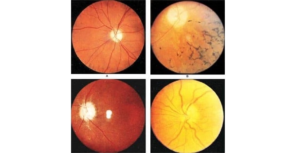
- Optic disc is greyish-white or white (chalky white) with clear margin.
- Shallow, saucer-shaped atrophic cupping due to degeneration of nerve fibres.
- Surrounding retina & retinal vessels are normal as the disease process is proximal (behind) the optic disc.
2. Consecutive optic atrophy
- Occurs following destruction of ganglionic cells secondary to degenerative or inflammatory lesions of choroid or retina.
Its common cause sre:
- retinitis pigmentosa
- diffuse chorioretinitis
- pathological myopia
- CRAO
- Glaucoma
- Disc is yellow-waxy in colour & the retinal vessels are attenuated.
3. Secondary optic atrophy
- Occurs after disc edema.
- Also c/d postpapilledematous optic atrophy
Its causes are:
- Papilloedema
- Optic neuritis (papillitis)
- Neuroretinitis
- Ischemic optic neuropathy
- Characterised by disappearance of vascularity of disc which causes increase in pallor of disc.
- Post-neuritic optic atrophy is a secondary optic atrophy which develops as a sequel to long standing papilloedema or papillitis.
4. Glaucomatous optic atrophy
- Occur in long stannding glaucoma i.e chronic open angle or closed angle glaucoma.
- There is glaucomatous cupping of optic disc.
- Abrupt margins of optic disc & normal retinal appearance.
5. Ischemic optic atrophy
- Due to disc ischemia caused by giant cell arteries, severe hemorrhage, severe anemia.
- Disc is pale with obliteration of retinal vessels.
6. Toxic optic atrophy
- Due to chronic retrobulbar neuritis secondary to toxic agents (toxic amblyopia)
Common causes are:
- tobacco
- ethyl alcohol
- methyl alcohol
- lead quinine
- chloroquinine

O/B of the pathological process, the optic atrophy can be divided into:
1. Ascending atrophy:
- Acsending optic atrophy (Wallerian degeneration) follows damage at retinal ganglion cell level.
- Nerve fiber degeneration progress (ascends) from the eye-ball towards the lateral geniculate body.
- So, consecutive optic atrophy is a type of ascending atrophy.
2. Descending atrophy:
- Descending (retrograde) atrophy.
- Occurs when degeneration proceeds from the region of optic tract, chiasma or retrobulbar portion of optic nerve towards optic disc.
- So, primary optic atrophy is a type of descending atrophy.
3. Cavernous (Schnable’s) optic atrophy:
- Mucoid degeneration of glia occurs.
Seen in:
- chronic simple glaucoma
- methyl alcohol poisoning
- high myopia
Exam Important
- Optic atrophy can be caused by Methyl alcohol poisoning.
- In optic atrophy pallor of the disc is an index of Loss of vascularty of the disc.
Don’t Forget to Solve all the previous Year Question asked on OPTIC ATROPHY
