Peritoneal cavity- lesseer sac & greater sac
PERITONEAL CAVITY
- Peritoneal cavity is the potential space b/w parietal & visceral layers & is filled with serous peritoneal fluid.
- Subdivided into: LESSEER SAC & GREATER SAC
LESSEER SAC (Omental bursa)
- Also c/d omental bursa or left subhepatic space or left posterior intraperitoneal space.
- It is a deep peritoneal space lying behind the stomach, lesser omentum & liver (caudate lobe).
- Site for abscess formation in posterior perforation of gastric ulcer and internal hernia through epiploic foramen.
- It is a closed space except for its communication on right side with greater sac through epiploic foramen.
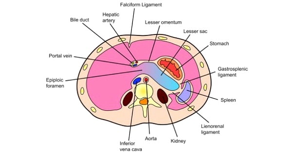
Boundaries of lesser sac are:
- Anterior wall: Caudate lobe of liver, stomach, Lesser omentum & 2nd layer of greater omentum
- Posterior wall: 3rd layer of greater omentum, & structurres forming stomach bed
- Right border: Right free margin of greater omentum & floor of epiploic foramen.
- Left border: Left free mergin of greater omentum; gastrosplenic & gastrophrenic ligaments
- Upper border: Reflection of peritoneum from esophagus to diaphragm.
- Lower border: Continuation of 2nd & 3rd layers of greater omentum.
Epiploic foramen (foramen of winslow or aditus to lesser sac) is a slit-like opening through which lesser sac communicates with greater sac.
- The foramen is about 3 cm in size and situated opposite the 12th thoracic vertebra.
Boundaries are:
- Anterior: Right free margin of lesser omentum (contains portal vein, hepatic artery proper & bile duct).
- Posterior: IVC, right suprarenal gland & T12 vertebra
- Superior: Caudate lobe of liver
- Inferior: 1st part of duodenum & horizontal part of hepoatic artery
- → A posterior gastric ulcer may perforate into lesser sac. The leaking fliud passes out through epiploic foramen to reach hepatorenal pouch.
- → Site for abscess formation in posterior perforation of gastric ulcer and internal hernia through epiploic foramen.

GREATER SAC
Greater sac is divided by line of attachment of transverse mesocolon & pelnic brim into 3 parts:
- Supracolic (above transverse mesocolon)
- Infracolic (below transverse mesocolon upto pelvic brim)
- Pelvic (below pelvic brim)
Exam Question
- The length of the epiploic foramen is 3 cm.
- Epiploic foramen provides communication between greater and lesser sacs.
- A posterior gastric ulcer may perforate into lesser sac.
- Omental bursa is site for abscess formation in posterior perforation of gastric ulcer and internal hernia through epiploic foramen.
Boundaries of lesser sac are:
- Anterior wall: Caudate lobe of liver, stomach, Lesser omentum & 2nd layer of greater omentum
- Posterior wall: 3rd layer of greater omentum, & structurres forming stomach bed
- Right border: Right free margin of greater omentum & floor of epiploic foramen.
- Left border: Left free mergin of greater omentum; gastrosplenic & gastrophrenic ligaments
- Upper border: Reflection of peritoneum from esophagus to diaphragm.
- Lower border: Continuation of 2nd & 3rd layers of greater omentum.
Don’t Forget to Solve all the previous Year Question asked on Peritoneal cavity- lesseer sac & greater sac