

RETINOPATHY OF PREMATURITY
RETINOPATHY OF PREMATURITY (ROM)
- Also k/a RETROLENTAL FIBROPLASIA.
- Affects retinal vasculature of premature babies.
- Retinal manifestations are noted some weeks after birth in premature infants who have been given high concentration of Oxygen.
- Confined to those with a birth weight of under 1.5 kg & gestational age 32 weeks.
- All babies weighing less than 1500 gm or having a gestational period of less than 32 weeks should be screened with indirect ophthalmoscopy for ROM, b/w 32 & 36 weeks post conception.
Important risk factors
1. Primary-
- Prematurity: Gestation age
- Birth weight< 1.5 kg
- Oxygen therapy: Excessive oxygen use
2. Other-
- Sepsis
- multiple blood transfusions
- Metabolic acidosis
Importnant signs
- Leukoplakia (white pupil)
- Nystagmus (abnormal eye movements)
- Strabismus (crossed eye)
- Severe nearsightedness (myopia)
Classification of ROP
I. O/B of severity, ROP divided into 5 stages:
- Stage 1: appearance of a thin, flat, white structure at the junction of vascularised retina posteriorly & avascular retina anteriorly
- Stage 2: the demarcation line develops into a pink ir white elevation (ridge) of thickened tissue.
- Stage 3: proliferation of vessels over the ridge & into vitreous (extravitreal fibrovascular proliferation)
- Stage 4: partial retinal detachment
4a- partial detachment with macular sparing
4b- partial detachment with macula involved
- Stage 5: total retinal detachment

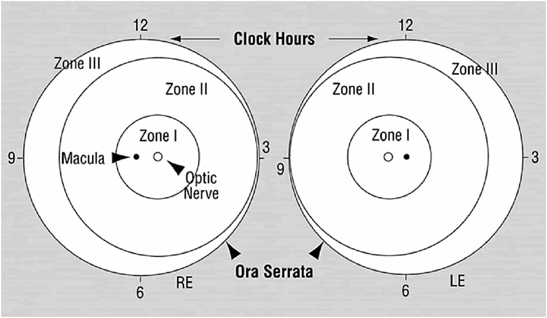
II. O/B of anatomical location:
- 3 zones are divided to describe the location of ROP.
- Location of border in zone 1 is the most severe disease & in zone 3 least.
i) Zone 1:
- Defined as a circle, the center of which is the disc.
- Radius of which is twice the distance of the disc of the fovea.
ii) Zone 2
- Doughnut- shaped region
- Extends from the anterior border of zone 1 to within one disc-diameter of the ora serrata nasally & the anatomic equator temporarally.
iii) Zone 3 encompasses the residual temporal retina.
PLUS DISEASE:
- As ROP progresses, more & more shunting occurs in the neovascular tissue at the retinal vascular-avascular junction.
This increased retinal vascular blood flow results in:
– dilation & tortuosity of the major retinal arteries & veins in the posterior pole- described as “plus disease”.
- Plus disease is the hallmark of rapidly progressive ROP
- Denoted by adding a plus sign after the number of the ROP stage.

MANAGEMENT
- Most of the cases (approx. 80%) of ROP resolve spontaneously.
- ROP is divided into threshold & prethreshold disease.
1. Threshold disease:
- Defined as a stage 3+ ROP in zones 1 or 2
- Occupying at least 5 contaguous clock-hours or eight noncontagious clock-hours of retina.
- Treatment of threshold disease is -Laser photocoagulation.
2. Prethreshold disease: divided into 2 types:
- High risk or Type I- treatment is Laser photocoagulation
- Low risk or Type II- treatment is Weekly or twice weekly observation.
| TYPE I Prethreshold | TYPE II Prethreshold |
| Zone I ROP (any stage)+
Zone II, Stage 3 Zone II, Stage2/3+ |
Zone I, Stage ½ –
Zone II, Stage 3- |
| Laser photocoagulation | Weekly or twice observation |
Exam Important
- Retinopathy of prematurity is commonly predisposed by Less gestation age.
- For preventing Retinopathy of Prematurity, concentration of oxygen should be 50-60%.
- Stage 3 of ROP shows a ridge with retinal fibrovascular proliferation and neovasularisation.
- Plus disease is the hallmark of rapidly progressive ROP.
Don’t Forget to Solve all the previous Year Question asked on RETINOPATHY OF PREMATURITY

