


SCAPHOID FRACTURE
SCAPHOID FRACTURE
- Fracture scaphoid accounts for 60% of carpal injuries.
- More common in young adults.
- Rare in children and in elderly people.
Mechanisms of Injury
- Two mechanisms are described:
1. Radial compression and dorsiflexion occurring at the wrist during a fall on an outstretched hand.
- Results in an undisplaced fracture.
2. Hyperextension occurring at the wrist during a fall on an outstretched hand.
- Results in displaced fracture scaphoid.
→ Undisplaced fractures are less complicated than displaced fractures.
Classifications
A. Based on Mayo classification:
- Fracture of the tuberosity
- Fracture of the distal body
- Fracture of the waist (m/c)
- Fracture of the proximal pole
- Osteochondral fracture
B. Based on Russe classification:
- Stable
- Unstable
Clinical signs that help in diagnosis
- Tenderness in the anatomical snuff box.
- Tenderness over the scaphoid tubercle
- Scaphoid compression test
Painful limitation of movements of wrist and thumb.


Radiological views taken to diagnose fracture scaphoid
- Standard PA view of the wrist (with wrist in dorsiflexion and ulnar deviation after making a fist)
- Standard lateral view.
- Radioulnar oblique view in midprone position.
- Stress views only if needed (Stress view opens up the fracture site).
Interpretation of the radiograph:
Scaphoid fracture is generally identified as:
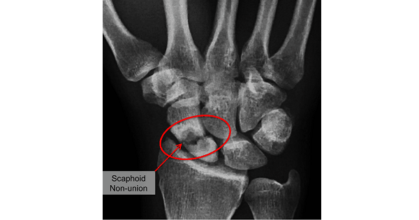
- clear lucent line across the bone.
- distinct break in continuity.
- A distinct sharp step.


Complications of Fracture Scaphoid
- Nonunion
- Avascular necrosis
Radiocarpal degenerative arthritis


Exam Important
- Most common site of scaphoid fracture is Waist.
- Fracture scaphoid is usually seen in Young active adult.
- Most common complication of scaphoid fracture is Avascular necrosis.
- Fracture of waist of scaphoid is most prone to develop Avanscular necrosis.
- Best radiological view for diagnosis of fracture scaphoid is Oblique.
Don’t Forget to Solve all the previous Year Question asked on SCAPHOID FRACTURE

