
Question
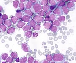
| A. | T(9,22) |
| B. |
T (15,17) |
| C. |
T (11, 14) |
| D. |
T(14, 18) |

|
Correct Answer � B Explanation |
 |
The image shows Auer cells
Auer rods, or Auer bodies, are cytoplasmic inclusions found in certain types of leukemic cells, most notably in acute myeloid leukemia (AML). They were first described by the American pathologist John Auer and are named after him.
Here are some key features of Auer rods:
Appearance: Auer rods are elongated, needle-like structures that can vary in size and number. They are often seen as red or pink staining structures in the cytoplasm of the cells when visualized with a light microscope and stained with common hematological stains like Wright or Giemsa stain
Composition: Auer rods are composed of crystallized or clumped granules containing enzymes such as myeloperoxidase, lysosomal enzymes, and other proteins related to granulocyte function.
Associated Conditions: Auer rods are typically associated with acute myeloid leukemia, particularly the myeloblastic types of AML (M1, M2, M3). They are considered pathognomonic for AML and are not typically seen in normal or reactive cells. In the M3 subtype, known as acute promyelocytic leukemia (APL), Auer rods may be seen in bundles, forming so-called “faggot cells.
Clinical Significance: The presence of Auer rods in leukemic cells is an important diagnostic feature for AML and provides information about the differentiation and nature of the leukemic blasts.
Potential Complications: The enzymes contained within Auer rods may contribute to disseminated intravascular coagulation (DIC), a severe and potentially life-threatening clotting disorder seen in some patients with AML.
Translocation and associated disorders
T(9,22): This translocation results in the Philadelphia chromosome, associated with chronic myeloid leukemia (CML) and some types of acute lymphoblastic leukemia (ALL).
T(15,17): This translocation is associated with acute promyelocytic leukemia (APL), a subtype of acute myeloid leukemia (AML).
T(11,14): This translocation is often seen in mantle cell lymphoma, a type of non-Hodgkin lymphoma.
T(14,18): This translocation is common in follicular lymphoma, another type of non-Hodgkin lymphoma.
