Question
| A. | Putty kidney |
| B. |
Pyelonephritis |
| C. |
Nephrocalcinosis |
| D. |
Staghorn calculus |

|
Correct Answer » A Explanation |
 |
Detailed Explanation:
Anatomical Role:
– The kidney filters blood, forming urine; chronic infections and inflammation can cause pathological changes evident on imaging.
Clinical Reasoning for the Correct Answer:
– The patient’s symptoms of abdominal pain, weight loss, and sterile pyuria suggest chronic renal pathology, often tuberculosis.
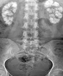
– The X-ray shows dense, diffusely calcified kidney resembling “putty,” characteristic of end-stage renal tuberculosis (putty kidney).
– Putty kidney results from granulomatous destruction, fibrosis, and dystrophic calcification of the renal parenchyma.
– Sterile pyuria (white blood cells without bacteria) is classic for renal tuberculosis as the causative mycobacteria are difficult to culture in routine bacterial cultures.
Why Option A is Correct:
– Putty kidney reflects end-stage renal tuberculosis with diffuse renal calcification.
– Clinical features match chronic TB: weight loss, abdominal pain, sterile pyuria.
– Radiology shows dense, diffuse renal calcification consistent with putty kidney.
Why Option B (Pyelonephritis) is Incorrect:
– Pyelonephritis presents acutely with fever, flank pain, and bacteriuria, not sterile pyuria.
– Radiographs rarely show dense diffuse calcifications; scarring may occur but not diffuse “putty”-like calcification.
– Routine urine cultures are positive in pyelonephritis.
Why Option C (Nephrocalcinosis) is Incorrect:
– Nephrocalcinosis is diffuse calcium deposition in the renal parenchyma, but typically symmetrical and due to metabolic causes like hyperparathyroidism or renal tubular acidosis.
– The clinical presentation lacks metabolic disturbance signs, and sterile pyuria is uncommon.
– Calcifications appear more granular rather than forming a dense “putty” pattern.
Why Option D (Staghorn Calculus) is Incorrect:
– Staghorn calculi are large renal pelvis stones shaped like the renal pelvis and calyces, visible as branching calcifications.
– Usually, bacterial infection (urease-positive) is present, and urine culture is positive.
– Calcification is localized to pelvis and calyces, not diffuse renal parenchymal calcification.
Differential Diagnoses
– Pyelonephritis: acute febrile illness, bacteriuria present.
– Nephrocalcinosis: metabolic causes, granular calcifications, symmetrical involvement.
– Staghorn calculus: branching calcifications localized in renal pelvis, positive urine cultures.
– Diagnostic Findings
– X-ray: Diffuse dense renal calcification (“putty kidney”).
– Urine analysis: sterile pyuria.
– Positive acid-fast bacilli (AFB) testing/culture confirms TB.