Question
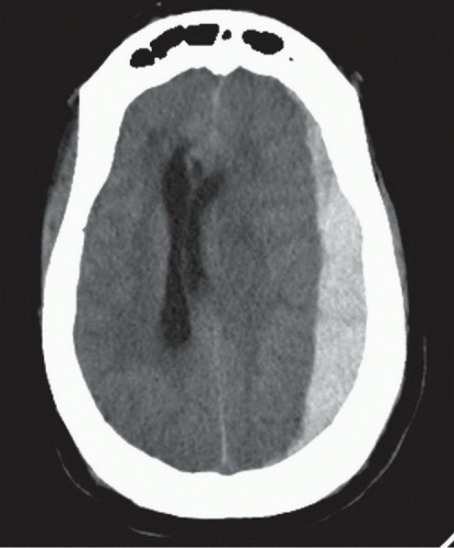
Which type of intracranial hemorrhage is most likely depicted in the image given below?
| A. |
Extradural hemorrhage
|
| B. |
Subarachnoid hemorrhage
|
| C. |
Subdural hematoma
|
| D. |
Intraparenchymal hemorrhage
|
Show Answer
|
Correct Answer » C
Explanation
|
|
-
The CT head image shows a crescent-shaped hyperdensity along the convexity of the cerebral hemisphere, located between the brain parenchyma and the skull.
-
The hemorrhage does not cross the midline but does extend across suture lines, which is characteristic of a subdural hematoma.
-
The red arrow is pointing to this crescentic collection, indicative of acute subdural bleeding.
Why Other Options Are Incorrect:
-
A. Extradural (epidural) hemorrhage:
-
Appears biconvex (lentiform) in shape.
-
Does not cross suture lines, but can cross the midline.
-
Often associated with temporal bone fractures and middle meningeal artery rupture.
-
Not seen here.
-
B. Subarachnoid hemorrhage (SAH):
-
Blood typically accumulates in the basal cisterns, sulci, and interhemispheric fissures.
-
Appears as hyperdensity within the CSF spaces.
-
Not the pattern seen in this image.
-
D. Intraparenchymal hemorrhage:
-
Presents as a localized hyperdense area within the brain parenchyma.
-
Does not show crescent shape.
-
Commonly due to hypertension, amyloid angiopathy, or vascular malformations.