
Question
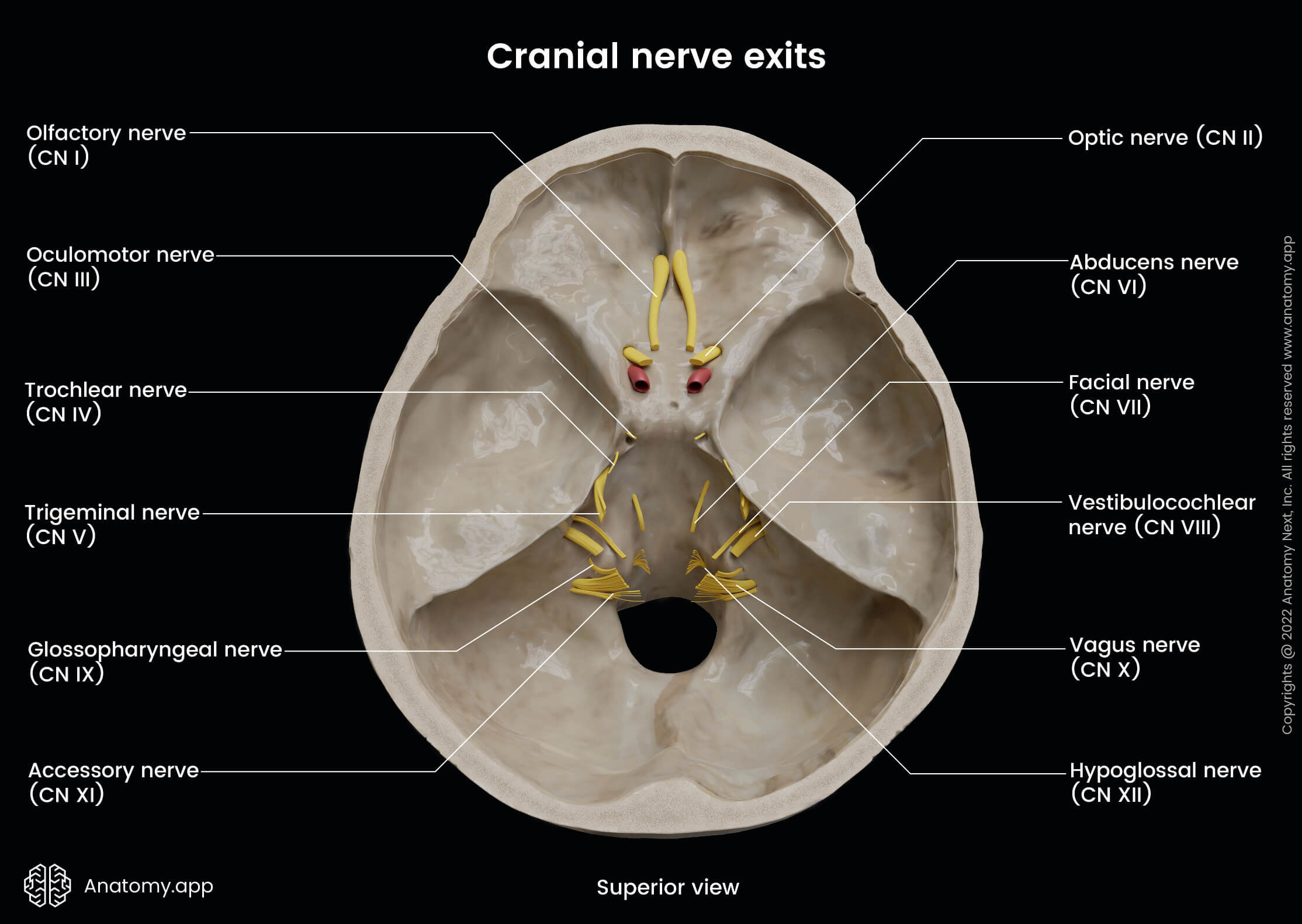
Which of the following cranial nerves is marked in the given image?

| A. | Oculomotor nerve |
| B. |
Abducens nerve |
| C. |
Trochlear nerve |
| D. |
Ophthalmic nerve |

|
Correct Answer � A Explanation |
 |
The nerve indicated by the arrow in the image is located at the superior orbital fissure and appears larger than the trochlear nerve. This positioning and size align with the oculomotor nerve (CN III), which passes through the superior orbital fissure and is responsible for motor innervation to most of the extraocular muscles.
The oculomotor nerve (CN III)is a prominent nerve passing through the superior orbital fissure and supplies:
- Superior rectus
- Inferior rectus
- Medial rectus
- Inferior oblique
- Levator palpebrae superioris (for eyelid elevation).
Its size and position match the structure pointed to in the image.
Functions: Motor innervation to most extraocular muscles and parasympathetic fibers to the sphincter pupillae and ciliary muscles.
Clinical Relevance:
- Lesions lead to ptosis, mydriasis, and inability to move the eye upward, downward, or medially.
- Common causes include aneurysms of the posterior communicating artery or cavernous sinus thrombosis.
Other Options:
Abducens nerve: The abducens nerve (CN VI) passes medially in the cavernous sinus and also through the superior orbital fissure. However, it is smaller and positioned more medially compared to the marked structure.
Trochlear nerve: The trochlear nerve (CN IV) is the smallest cranial nerve and lies laterally within the cavernous sinus before entering the orbit. It supplies the superior oblique muscle. Its size and position do not match the marked structure.
Ophthalmic nerve: The ophthalmic nerve (V1) is a branch of the trigeminal nerve. It passes through the superior orbital fissure and divides into frontal, lacrimal, and nasociliary branches. This nerve is significantly larger and does not correspond to the marked structure.
