
PEPTIC ULCER (GASTRIC AND DUODENAL ULCER)
Which one is not associated with peptic ulcer –
| A | Smoking cigarrete | |
| B | Zollinger Ellison’s synd | |
| C |
Plummer vinsons synd |
|
| D |
Cirrhosis |
Which one is not associated with peptic ulcer –
| A |
Smoking cigarrete |
|
| B |
Zollinger Ellison’s synd |
|
| C |
Plummer vinsons synd |
|
| D |
Cirrhosis |
Ans is ‘c’ ie Plummer-Vinson syndrome
- Conditions asso. with peptic ulcer have already been described.
- Plummer Vinson Paterson Kelly syndrome consists of :‑
a) Iron deficiency anemia
b) Dysphagia [d/t fibrous esophageal web at its upper end *]
c) atrophic oral mucosa, glossitis
d) brittle spoon shaped finger nails.
- Typically pts are Woman over 40 yrs of age.
- Its a Pre-malignant condition* for both oral cavity & esophagus.
Commonest site of peptic ulcer is –
| A | Ist part of duodenum | |
| B | IInd part of duodenum | |
| C |
Distal 1/3 of stomach |
|
| D |
Pylorus of the stomach |
Ans. is ‘a’ i.e., 1st part of Duodenum
- Duodenum, 1st portion
- Stomach, usually antrum
- At the gastro – esophageal junction, in the setting of gastroesophageal reflux or Barrett’s esophagus
- Within the margins of a gastrojejunostomy
- In the duodenum, stomach or jejunum of patients with Zollinger-Ellison Syndrome
- Within or adjacent to Meckels diverticulum that contains ectopic gastric mucosa
In gastric outlet obstruction in a peptic ulcer patient, the site of obstruction is most likely to be-
| A | Antrum | |
| B |
Duodenum |
|
| C |
Pylorus |
|
| D |
All |
Ans. is ‘b’ i.e., Duodenum
- Bailey writes – “The term ‘pyloric stenosis’ is a misnomer. The stenosis is seldom at the pylorus. Commonly, when the condition is due to underlying peptic ulcer disease, the stenosis is found in the first part of the duodenum, the most common site for a peptic ulcer.”
- Also know
- There are 2 common causes of gastric outlet obstruction
– pyloric stenosis secondary to peptic ulcer
– gastric cancer
– Earlier peptic ulcer was the most common cause, but now the most common cause of gastric outlet obstruction is —> gastric cancer.
A pt. of peptic ulcer disease. When investigated endoscopically showed chronic antral gastritis. Which of the following dye will be able to stain the specimen –
| A | PAS | |
| B |
Zeil-Neilson stain |
|
| C |
Gram stain |
|
| D |
Warthin-Starry stain |
Ans. is ‘d’ ie. Warthin starry stain
Most cases of chronic gastritis are one of the two types –
Type A – Autoimmune type associated with pernicious anemia, it primarily involves the body and fundus. Type B – associated with H. pylori infection, it primarily involves the antrum.
Mnemonic
Type A – Autoimmune
Type B – Bacteria i.e. H. pylori
- In this pt. having chronic antral gastritis (type B) we will use a dye which will help stain H. pylori.
- Dye used for H. pylori is Warthin starry stain (or Giemsa or genta silver stain)*
Some other imp stains
Zeil Nelson* —> i i ()bacterium tuberculosis
Albert stain* —> Corynebacterium diptheria Oil red 0 stain* –p Fats
PAS* —> Glycogen
Prussian blue* -> Iron
Congo red * –> Amyloid
Von Kossa* —> Calcium
| A |
i.v. fluids |
|
| B |
Drainage of paracolic gutter |
|
| C |
Immediate surgery |
|
| D |
a and c |
Ans ‘a’ fluids; ‘c’ immediate surgery
Management of Peptic ulcer perforation
Nasogastric tube: whenever a perforated ulcer is suspected, the first step is to pass a nasogastric tube and empty the stomach to reduce further contamination of the peritoneal cavity.
Intravenous crystalloid: The patient is resuscitated aggressively by administration of intravenous crystalloid.
– Intravenous broad-spectrum antibiotics
Surgery:
Surgery is mostly indicated, although occasionally nonsurgical treatment can be used in stable patients without peritonitis, and in whom radiologic studies document a sealed perforation.
Surgery whether laparoscopy or laparotomy involves two components:
i) Thorough peritoneal toilet
to remove all the fluid and food debris
drain is not indicated
ii) Management of perforation
For duodenal ulcer perforation
The most frequently performed operation for a perforated duodenal ulcer is simple closure with an omental onlay reinforcement or patch.
This is combined with postoperative H. pylori eradication (antibiotics + antisecretory agents)
– Insertion of a nasoenteric or jejunal feeding tube should be considered
[Note that this current practice is in marked contrast to the prior recommendation suggesting the addition of a concomitant acid-reduction procedure to ulcer closure.]
For gastric ulcer perforation
All perforated gastric ulcers are best treated by distal gastric resection with or without a truncal vagotomy (Truncal vagotomy can be added for type II & III gastric ulcers. Type I and IV gastric ulcers do not need acid reducing procedures as they are associated with hyposecretion). Ulcer is removed along with gastric resection as it carries a risk of malignancy (cf: duodenal ulcer has no risk of cancer). If ulcer is left behind, biopsy is always done.
Other procedures used for gastric ulcers are
– patch closure with biopsy – wedge excision and closure
| A |
Posteriorly perforated ulcers are always managed conservatively |
|
| B |
Anterior ulcers bleed more commonly Increased acid production is prerequisite for gastric ulcer |
|
| C |
Anti-H. pylori drugs must be included in the treatment regimen |
|
| D |
H. pylori is known to increase the incidence of gastric malignancy |
Ans is ‘d’ (H. pylori is known to increase the incidence of gastric malignancy)
Surgery is almost always indicated for perforated ulcers, although occasionally nonsurgical treatment can be used in stable patients without peritonitis, and in whom radiologic studies document a sealed perforation.
Though increased acid production is associated with duodenal ulcers, its not so with gastric ulcers. Gastric ulcers type I and type IV are associated with decreased acid secretion. (Gastric type II and type III are associated with acid hypersecretion)
Anti-H.pylori drugs are included in those pts. of peptic ulcer ds. who test positive for H. pylori.
Commonest site of peptic ulcer perforation ‑
| A | Anterior aspect of the first part of duodenum | |
| B | Posterior aspect of the 1st part of duodenum | |
| C |
Greater curvature of the stomach |
|
| D |
Lesser curvature of the stomach |
Ans. is ‘a’ i.eAnterior aspect of the first part of duodenum
Percentage of patients with perforated peptic ulcer who show free gas under the diaphragm –
| A |
100% |
|
| B |
75% |
|
| C |
50% |
|
| D |
90% |
Ans. is ‘b’ i.e. 75%
Sutton writes – ”The demonstration of pneumoperitoneum on plain film following a perforated viscus is, however, not invariable, and most series show that in only 75-80% of perforations is free gas demonstrable. A number of reasons for this have been suggested, including sealing of the perforation, lack of gas at the site of perforation, or adhesions around the site of perforation. However, radiographic technique is also important : a pneumoperitoneum can be detected in 76% of cases using an erect film only, but when a left lateral decubitus projection is included, a pneumoperitoneum is demonstrated in nearly 90% of cases.”
Commonest operation done for peptic ulcer with gastric outlet obstruction is –
| A |
Truncal vagotomy with pyloroplasty |
|
| B |
Highly selective vagotomy with pyloroplasty |
|
| C |
Truncal vagotomy with gastrojejunostomy |
|
| D |
Gastrojejunostomy |
Ans. is ‘c’ i.e., Truncal Vagotomy with gastrojejunostomy
Gastric outlet obstruction due to peptic ulcer can develop either acutely or chronically.
Acute obstruction
Is due to edema and inflammation..
Acute gastric outlet obstruction is treated by nasogastric suction, rehydration, and intravenous
administration of antisecretory agents. Acute obstruction due to pyloric inflammation resolves with
supportive measures within 72 hours.
Chronic obstruction
Repeated episodes of ulceration can lead to pyloric scarring and a fixed stenosis with chronic gastric
outlet obstruction.
Surgery for duodenal ulcer:
– Truncal vagotomty with antrectomy (surgery of choice)
– Truncal vagotomy with gastrojejunostomy
– Surgery for gastric ulcer
– Distal gastrectomy
| A | Perforation | |
| B |
Hemorrhage |
|
| C |
Pyloric stenosis |
|
| D |
Maliganancy |
Ans. is ‘b’ i.e., Hemorrhage
Investigation of choice in peptic ulcer perforation is ‑
| A |
USG |
|
| B |
X-Ray abdomen |
|
| C |
Paracentesis |
|
| D |
CT scan |
Ans. is ‘b’ i.e., X-ray abdomen
In the Forrest classification for bleeding peptic ulcer with a visible vessel or pigmented protuberance is classified as –
| A |
Et |
|
| B |
FII a |
|
| C |
FII b |
|
| D |
FII c |
Ans. is ‘b’ i.e., FII a
Forrest Classification
Peptic ulcer disease is the most common cause of upper gastrointestinal bleeding. The endoscopic finding can give valuable information about the risk of rebleeding after hospital admission. Endoscopic signs indicating risk of rebleeding were classified in 1974 by Forrest et al. The classification is as follows –
|
Forrest class |
Type of lesion |
Risk of rebleeding if untreated |
|
IA |
Arterial spurting bleeding |
100% |
|
IB |
Arterial oozing bleeding |
55% |
|
IIA |
Visible vessel |
43% |
|
IIB |
Sentinel clot |
22% |
|
IIC |
Hematin covered flat spot |
10% |
|
III |
No stigmata of hemorrhage |
5% |
A 20 year old male has presented with repeated episodes of hematemesis. There is no history of jaundice or liver decompensation. On examination the significant findings include splenomegaly (8 cms below costal margin), and presence of esophageal varices. There is no ascites or peptic ulceration. The liver function tests are normal. The most likely diagnosis is-
| A | Extahepatic portal venous obstruction | |
| B | Non cirrhotic portal fibrosis | |
| C |
Cirrhosis |
|
| D |
Hepatic venous outflow tract obstruction |
Ans. is ‘b’ i.e., Non cirrhotic portal fibrosis
Splenomegaly, variceal bleeding and absence of any abnormality in the liver in a young adult suggests the diagnosis of Non cirrhotic portal fibrosis (NCPF).
Remember extrahapatic portal venous obstruction also presents with similar features but the important features to distinguish b/w these two conditions is, age of the patient
Age of presentation: ‑
EHPVO —> Children (1st to 2nd decade) NCPF Young adult (2nd to 3rd decade)
|
Comparison |
b/w NCPF and EHPVO Difference |
|
Similarities |
|
|
Massive variceal bleeding |
Age of presentation |
|
Moderate to massive Splenomegaly |
EHPVO —> Children |
|
Normal liver on histology |
NCPF —> Young adult |
|
Absence of Ascites |
Site of obstruction in the portal venous system |
|
Normal liver function |
EHPVO —> Main and 1st order branches of portal vein |
|
|
NCPF —> Smaller 3rd and 4th order branches of portal vein are involved |
A 45-year-old gentleman has undergone truncal vagotomy and pyloroplasty for bleeding duodenal ulcer seven years ago. Now he has intractable recurrent symptoms of peptic ulcer. All of the following suggest the diagnosis of Zollinger Ellison syndrome, except:
| A | Basal acid output of 15 meq/hour | |
| B | Serum gastrin value of 500 pg/ml | |
| C |
Ulcers in proximal jejunum and lower end of esophagus |
|
| D |
Serum gastrin value of 200 pg/ml with secretin stimulation |
Ans. is ‘d’ i.e. Serum gastrin value of 200 pg/ml with secretin stimulation
Diagnosis of ZES is made by secretin provocative test when serum gastrin value increases 200 pg/mL within 15 minutes [ Look closely at the option (d). It says serum gastrin value of 200 pg/mL with secretin stimulation. It mentions the absolute serum gastrin level, not the increase in level]
Zollinger Ellison syndrome
- ZES is caused by gastrin secreting gut neuroendocrine tumors (gastrinomas), which result in hypergastrinemia and acid hypersecretion.
- Clinical situations that create suspicion for ZES are
– ulcers in unusual locations, ulcers in second part of duodenum & beyond (includes the option c) ulcers refractory to standard medical therapy
– ulcer recurrance after acid reducing surgery.
– ulcers presenting with frank complications (bleeding, perforation and obstruction)
– ulcers in the absence of H. pylori or NSAID ingestion.
– giant ulcers (> 2 cm)
– multiple duodenal ulcers
– ulcers associated with diarrhoea
– ulcer patient with hypercalcemia or family h/o ulcers.
– ulcers with severe esophagitis.
– ulcers associated with prominent gastric folds
– ulcers with findings s/o MEN-1 (endocrinopathy, family h/o ulcers or endocrinopathy, nephrolithiasis)
Diagnosis of ZES (gastrinoma)
1) The first step in evaluation of a patient suspected of having ZES or gastrinoma is to obtain as fasting gastrin
level.
All gastrinoma patients have a fasting gastrin level > 150 pg/mL.
2) The next step is to assess acid secretion by measuring basal acid output (BAO) which should be Z 15 meq/h (normal < 4 meq/h)
[This is done to rule out hypochlorhydria which is much more common cause of hypergastrinemia than
gastrinoma]
A gastric pH of > 3.0 implies hypochlorhydria and excludes gastrinoma.
3) Gastrin Provocative tests
– In a pt. with pH < 2.0 or BAO . 15 meq/hr and fasting gastrin level > 1000 pg/mL, the diagnosis of ZES is confirmed.
– With pH < 2.0 or BAO 15 meq/L and lower gastrin levels (150-1000 pg/mL), a secretin stimulation test is perforfmed to distinguish ZES from other causes of hypergastrinemia. A rise of 200pg/mL of serum gastrin level within 15 min of secretin injection, confirms ZES.
The hernia which often simulates a peptic ulcer is ‑
| A | Umbilical hernia | |
| B | Fatty hernia of the linea alba | |
| C |
Incisional hernia |
|
| D |
Inguinal hernia |
Ans. is ‘b’ i.e., Fatty hernia of the linea alba
Intractable peptic ulceration with renal stones occurs in –
| A |
Zollinger Ellison syndrome |
|
| B |
Parathyroid adenoma |
|
| C |
Milk, alkali syndrome |
|
| D |
MEN I syndromeVicryl |
Ans. is ‘d’ i.e., MEN 1 syndrome
Peptic ulcer is associated with all except one:
| A |
Cirrhosi |
|
| B |
Zollinger Ellisons syndrome |
|
| C |
Primary hyperparathyroidism |
|
| D |
Pernicious anemia |
Answer is D (Pernicious anemia):
Pernicious anemia is characterized by autoimmune destruction of parietal cells leading to hypochondria (J, acid production). It is not associated with peptic ulcer disease.
Aetiological Factors in peptic ulceration
Primary Aetiologcial Factors (selected)
- H. pylori infection
- NSAID’s and other drugs eg. steroids
- Stress – Head injury / Burns
- Bile Reflux
- Smoking /Alcohol use
- Genetic Factors
Associated Factors / Conditions (selected)
- Endocrinal Disorders – MEN
– Primary Hyperparathyroidism
– Zollinger Ellison Syndrome
– Cushing’s syndrome
- Cirrhosise
- Chronic Renal Failure
- Chronic Pulmonary Disease
- Chronic Paracreatic Disease
- Alpha 1 Antitrypsin deficiency
| A | Smoking cigarette | |
| B |
Zollinger Ellison syndrome |
|
| C |
Plummer Vinsons syndrome |
|
| D |
Cirrhosis |
The answer is C (Plummer Vinson Syndrome):
Common causes include:
Helicobacter pylori and non-steroidal anti-inflammatory drugs (NSAIDs).
tobacco smoking
stress due to serious illness
Behcet disease
Zollinger-Ellison syndrome
Crohn disease
liver cirrhosis.
Older people are more sensitive to the ulcer-causing effects of NSAIDs.
Which of the following statements about peptic ulcer disease is true :
| A |
Helicobacter pylori eradication increases the likelihood of occurrence of complications |
|
| B |
The incidence of complications has remained unchanged |
|
| C |
The incidence of Helicobacter pylori reinfection in India is very low. |
|
| D |
Helicobacter pylori eradication does not alter the recurrence ratio. |
Answer is B (The incidence of complications has remained unchanged)
- H. pylori eradication decreases the likelihood of occurrence of complications.
- The incidence of H.pylori is > 80% in most developing countries, and so the chances of reinfection are also high.
- H. pylori eradication reduced the chances of recurrence
- ‘After successful eradication of H.pylori with antibiotics, ulcer recurrence rates are reduced dramatically to 5-20% at 1 year – CMDT 2003/571
- Option ‘b’ ‘The incidence of complications has remained unchanged’ is thus the answer of exclusion !!
| A |
1st part of Duodenum |
|
| B |
2nd part of duodenum |
|
| C |
Distal 1/3 of stomach |
|
| D |
Pylorus of the stomach |
Answer is A (1st part of Duodenum)
MC site: ₋₋ 1st part of duodenum (overall MC site for peptic ulcer)
Sites in order of decreasing frequency :
- Duodenum, 1st portion
- Stomach, usually antrum
- At the gastro-esophageal junction, in the setting of gastroesophageal reflux
- Within the margins of a gastrojejunostomy
- In the duodenum, stomach or jejunum of patients with Zollinger- Ellison Syndrome
- Within or adjacent to a Meckel’s diverticulum that contains ectopic gastric mucosa
Peptic ulcers are usually solitary lesions less than 4 cm in diameter
| A |
Gastric resection |
|
| B |
Vagotomy + drainage |
|
| C |
Vagotomy + Antrectomy |
|
| D |
Highly selective vagotomy |
Answer is C (Vagotomy + Antrectomy)
Repeat Vagotomy with Antrectomy is associated with lowest recurrence rates.
In gastric outlet obstruction in a peptic ulcer patient the site of obstruction is most likely to be :
| A |
Antrum |
|
| B |
Duodenum |
|
| C |
Pylorus |
|
| D |
Pyloric canal |
Answer is B (Duodenum)
Gastric outlet obstruction in a patient with peptic ulcer disease is suggestive of pyloric stenosis. The term pyloric stenosis is normally a misnomer. The stenosis is seldom at the pylorus.
Commonly the stenosis is found in the first part of the duodenum, the most common site for a peptic ulcer.
There is no history of jaundice or liver decompensation.
On examination there, significant findings include splenomegaly (8 ems below costal margin), and the presence of esophageal varices.
There is no ascites or peptic ulceration.
The liver function tests are normal.
The most likely diagnosis is
| A |
Extahepatic portal venous obstruction |
|
| B |
Non cirrhotic portal fibrosis |
|
| C |
Cirrhosis |
|
| D |
Hepatic venous outflow tract obstruction |
The answer is B (Noncirrhotic portal fibrosis) :
Features of portal hypertension in an adult with large splenomegaly (8cm below costal margin) with no history of jaundice or liver decompensation suggests a diagnosis of Non-cirrhotic portal hypertension.
Non-cirrhotic portal hypertension encompasses two distinct pathological conditions that present with similar clinical features.
The distinction between the two conditions should ideally be made investigations are similar in presentation makes clinical criteria unreliable.
Nevertheless, if we do have to choose one single best option the following clues may be helpful.
NCPF
- NCPF presents in young adults most commonly during the third and fourth decade
- Mode of presentation
- The onset of symptoms is gradual
- Mass in left upper quadrant (splenomegaly) is about 4 times more common in NCPF than EHPH
Extrahepatic portal hypertension
- EPH may present in two age groups children (Ist & 2nd decade) (congenital malformations) Adults (4th & 5th decade) (thrombotic event)
- The onset is gradual in children where the cause is a congenital malformation
- The onset is acute in adults where the cause is usually a thrombotic even
- Mass in left upper quadrant (splenomegaly) is four times less common in patients with EHPH
Thus a young adult presenting with a gradually progressive cause and a moderate to massive splenomegaly suggests the diagnosis of noncirrhotic portal fibrosis.
March 2008
| A |
Usually multiple |
|
| B |
More common in the stomach |
|
| C |
Round to oval, sharply punched out defect |
|
| D |
Size and location helps to differentiate between benign and malignant ulcer |
Ans. C: Round to oval, sharply punched out defect
Peptic ulcer are four times more common in the proximal duodenum than in the stomach.
Peptic ulcers are solitary in more than 80% of the patients.
Peptic ulcers are sharply punched out with straight walls and a flat base which is smooth. The adjoining mucosa is flat and the rugal folds extend to the margin of the ulcer and some reddening of the adjoining mucosa may indicate an associated gastritis. The gastric mucosa is often flattened due to the presence of chronic gastritis with atrophy. Acute ulcers may show edema and chronic ulcers show varying degrees of underlying fibrosis. The depth of the ulcer is variable with some showing only mucosal involvement in which case they are called erosions in contrast to deeply penetrating ulcers which involve submucosa and deeper layers. Occasionally evidence of active bleeding may be seen, and deep ulcers may perforate. Benign gastric ulcers are most commonly found along the lesser curvature near the interface of the body and antrum.
Size and location of the ulcer do not differentiate benign and malignant ulcers. However the gross appearance of chronic peptic ulcer is virtually diagnostic.
Malignant transformation of the peptic ulcer is very rare.
September 2012
| A | Clarithyromycin | |
| B |
Amoxycillin |
|
| C |
Ciprofloxacin |
|
| D |
Proton pump inhibitor |
Ans. C i.e. Ciprofloxacin
H. pylori
Its eradication drastically reduces ulcer recurrence in patients with duodenal or gastric ulcers.
- 7-day triple therapies with a proton pump inhibitor (PPI) + two antibiotics given BID are currently recommended first-line for H. pylori eradication e.g. lansoprazole (or alternate PPI) + clarithromycin + either metronidazole or amoxicillin
- A dose of clarithromycin 250mg p.o. BID is preferred when using in combination with a PPI and metronidazole; however, the 500mg p.o. BID dose is recommended in combination with a PPI + amoxicillin.
- Maintenance acid suppression therapy is not necessary following H. pylori eradication except in high-risk patients (e.g. severe GI bleed; refractory ulcer disease).
- Ranitidine Bismuth Citrate or RBC is a new agent useful in H. pylori eradication regimens.
March 2009
| A | Omeprazole | |
| B |
Ranitidine |
|
| C |
Misoprostol |
|
| D |
a and c |
Ans. A: Omeprazole, C: Misoprostol
Drugs for peptic ulcer and GERD with safety uncertain/unsafe profile in pregnancy include:
- Cimetidine
- Omeprazole, pantoprazole, lansoprazole
- Cisapride, mosapride
Safer drugs in pregnancy for peptic ulcer/GERD are:
- Ranitidine, famotidine
September 2006, March 2013(a)
| A | NSAID ingestion | |
| B |
Smoking |
|
| C |
H. pylori |
|
| D |
Genetic factors |
Ans. C. H.pylori
Helicobacter pylori is a Gram-negative, microaerophilic bacterium that can inhabit various areas of the stomach and duodenum.
It causes a chronic low-level inflammation of the stomach lining and is strongly linked to the development of duodenal and gastric ulcers and stomach cancer.
Over 80% of individuals infected with the bacterium are asymptomatic.
The most reliable method for detecting H. pylori infection is a biopsy check during endoscopy with a rapid urease test, histological examination, and microbial culture.
The standard first-line therapy is a one week “triple therapy”
March 2007
| A | Truncal vagotomy with gastrojejunostomy | |
| B |
Truncal vagotomy with gastrojejunostomy |
|
| C |
Highly selective vagotomy with pyloroplasty |
|
| D |
Gastrojejunostomy |
Ans. A: Truncal vagotomy with gastrojejunostomy
The diagnosis is typically made by X-ray, which will show an absent air bubble in the stomach, or by CT scan.
Causes of gastric outlet obstruction include peptic ulcer disease; tumours of the stomach, including adenocarcinoma (and its linitis plastica variant), lymphoma, and gastrointestinal stromal tumours; infections, such as tuberculosis; and infiltrative diseases, such as amyloidosis.
Metabolic alkalosis may develop as a result of loss of acid.
Traditionally severe cases of Gastric outlet obstruction are treated usually by a gastroenterostomy rather than a pyloroplasty. Truncal vagotomy leads to reduction of the maximal acid output by 50% as vagus nerves are sectioned which critically controls the secretion of gastric acid.
| A | Umbilical hernia | |
| B |
Incisional hernia |
|
| C |
Strangulated hernia |
|
| D |
Fatty hernia of linea alba |
Ans. D: Fatty hernia of the linea alba
Pain in the case of epigastric hernia, also known as a fatty hernia of the linea alba, may simulate peptic ulcer.
Epigastric hernia:
- Located between the xiphoid process and umbilicus, usually in the midline
- More common in males
- Pain due to the incarceration of the preperitoneal fat
- Pain simulates peptic ulcer
- No sac
| A |
Clopidogrel |
|
| B |
NSAID |
|
| C |
Mycophenolate mofetil |
|
| D |
Propylthiouracil |
Ans. is ‘d’ i.e., Propylthiouracil
Drug/Toxin causing peptic ulcer disease:
Bisphosphonates
Chemotherapy
Clopidogrel
Crack cocaine
Glucocorticoids (when combined with NSAIDs)
Mycophenolate mofetil
Potassium chloride
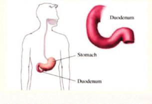
| A |
1st part. |
|
| B |
2nd part. |
|
| C |
3rd part. |
|
| D |
4th part. |
Organ shown in the picture above represents duodenum.
Peptic ulcer
o Ulcers are defined histologically as a breach in the mucosa of the alimentry tract that extends through the
mucularis mucasea into the submucosa or deep.
MC site: 1stpart of Duodenum (overall me site for peptic
ulcer)
o Peptic ulcers are chronic , most often solitary lesions that occur in any protion of the gastrointestinal tract exposed
to the aggressive action of acid / peptic juice .
Peptic ulcers located in the following sits, in order of decreasing frequency :
- Duodenum, 1′ portion
- Stomach ,usally antrum
- At the gastro – esophageal junction in the setting of gastroesphageal reflux or Barrett’s esophagus.
- Within the margins of a gastrojejunostomy.
- In the duodenum, stomach or jeunum of patients with Zollinger -Ellison Syndrome.
- Within or adiacent to Meckels diverticulum that contains ectopic gastric mucosa.
Most common site of peptic ulcer in duodenum ‑
| A | Pt part | |
| B |
2nd part |
|
| C |
3rd part |
|
| D |
41h part |
Ans. is ‘a’ i.e., lst part
Peptic ulcer
- Ulcers are defined histologically as a breach in the mucosa of the alimentry tract that extends through the mucularis mucasea into the submucosa or deep.
- Peptic ulcers are chronic , most often solitary lesions that occur in any protion of the gastrointestinal tract exposed to the aggressive action of acid / peptic juice .
Peptic ulcers located in the following sits, in order of decreasing frequency :
- Duodenum, ls`portion
- Stomach ,usally antrum
- At the gastro – esophageal junction in the setting of gastroesphageal reflux or Barrett’s esophagus.
- Within the margins of a gastrojejunostomy.
- In the duodenum, stomach or jeunum of patients with Zollinger -Ellison Syndrome.
- Within or adiacent to Meckels diverticulum that contains ectopic gastric mucosa.
