
PULMONARY EMBOLISM
PULMONARY EMBOLISM

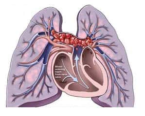

- Most common cause of preventable hospital death.
RISK FACTORS:
- More than 95% results from deep vein thrombosis.
Risk of blood clots increased by:
- Cancer
- Prolonged bed rest
- Smoking
- Stroke
- Large veins of lower limb
- Certain genetic conditions
- Estrogen-based medication
- Pregnancy
- Obesity
- After certain surgery
SYMPTOMS:
Most PE remains clinically silent.
- Dyspnea
- Pain
- Haemoptysis
- Tachypnea – Due to activation of juxtapulmonary-capillary receptors.
- Chest pain – “Pleuritic” nature (worsened by breathing).
- Cyanosis.
More severe signs:
- Attributes to about 15% of all sudden death cases.
- Collapse happens due to circulatory instability caused by decreased blood flow to lungs & left heart.
Saddle emboli:
- Large emboli block pulmonary artery bifurcation area resulting in saddle emboli presentation.
Infarction:
- Only 10%PEresults in infarction.
- Infarction will be wedge-shaped hemorrhagic type.
DIAGNOSIS:
- Computed tomography (CT) pulmonary angiogram (Best method).
Ventilation-perfusion scan:
- Gold standard.
- Findings – Perfusion segmental defect with normal lung scan & radiography.
- D-dimer test – Most sensitive test.
ECG findings:


- Sinus tachycardia – Most frequent ECG finding in acute pulmonary embolism.
Pattern –
- S1Q3T3 (Ie., Large S wave in lead I, Q wave in lead III, & inverted T wave in lead III).
- Also referred as “McGinn-White Sign”.
- Reveals right ventricular dilatation & tricuspid regurgitation.
Radiological findings:
Hamptons’ hump:
- Classic radiographic finding.
- Appears as peripheral wedge-shaped opacity on chest x rays.
Westermark’s sign –
A focal area of oligemia due to vasoconstriction distal to embolus.
Palla’s sign –
- Enlarged right descending pulmonary artery.
TREATMENT:
Thrombolytic therapy:
- Done in patients presenting with hemodynamic compromise during conventional therapy with heparin.
- Also with embolism associated with intracavitary right-heart thrombi.
Note:
- The anticoagulant effect of heparin in pulmonary embolism would be most appropriately assessed by Activated partial thromboplastin time.
Exam Important
- Best investigation when there is clinical suspicion of pulmonary embolism in a patient is Multi detector CT angiography
- Hampton’s hump is seen in chest X-ray in Pulmonary embolism.
- Test results that confirm or exclude pulmonary embolism are Perfusion scan & CT pulmonary angiogram
- Hampton’s hump, Westermark sign & Fleischner sign are radiological sign present in a young lady with symptoms suggestive of pulmonary embolism.
- The anticoagulant effect of heparin in pulmonary embolism would be most appropriately assessed by Activated partial thromboplastin time.
- Treatment of choice for acute pulmonary embolism with right ventricular hypokinesis and compromised cardiac output but normal blood pressure is Thrombolytic agent.
- Obesity, Malignancy & Protein S deficiency may predispose to pulmonary embolism
- In acute pulmonary embolism, Sinus tachycardia is the most frequent ECG finding
- Activation of juxtapulmonary-capillary receptors during pulmonary embolism causes Tachypnea
- The most common source of pulmonary embolism is Large veins of lower limb.
- D-dimer is the most sensitive test for Pulmonary embolism
- In pulmonary embolism, findings in perfusion scan are perfusion segmental defect with normal lung scan & radiography.
- Best method to diagnose pulmonary embolism Pulmonary angiography
- Gold standard for diagnosing pulmonary embolism Ventilation-perfusion scan
- The sequence of symptoms in pulmonary embolism is Dyspnea, pain, haemoptysis
- In pulmonary embolism, fibrinolytic therapy is responsible for risk of haemorrhage
- A patient undergoing surgery suddenly develops hypotension, the monitor shows that end-tidal CO2 has decreased abruptly by 155 mm Hg. Diagnosis – Pulmonary embolism
- The most common cause of preventable Hospital Death is Pulmonary embolism
- ECG pattern seen in pulmonary embolism is S1Q3T3.
Don’t Forget to Solve all the previous Year Question asked on PULMONARY EMBOLISM
Click Here to Start Quiz
Click Here to Start Quiz

