
Pulmonary Embolism
Best investigation when there is clinical suspicion of pulmonary embolism in a patient is
| A |
D – Dimer assay |
|
| B |
Multi detector CT angiography |
|
| C |
Doppler ultrasound |
|
| D |
Catheter angiography |
Best investigation when there is clinical suspicion of pulmonary embolism in a patient is
| A |
D – Dimer assay |
|
| B |
Multi detector CT angiography |
|
| C |
Doppler ultrasound |
|
| D |
Catheter angiography |
Multidetector CT angiography [Ref: Harrison 17th/e p. 1653-1654 & 16th/e p. 1563]
Diagnosis of pulmonary embolism ?
- A low likelhood case on clinical assessment should .first undergo D – Dimer assays. Only, the patients, who are positive on D – Dimer should undergo. further evaluation (imaging tests).
- On the other hand if the patient has high clinical likelihood on clinical assessment he should straightaway undergo imaging tests.
- The first or the principal imaging test for pulmonary embolism is computed tomography (CT) of the chest with intraveous contrast.
- These multidetector row spiral CT’s have replaced ventilation perfusion scan as the initial imaging modality in 1 pulmonary embolism.
- V/0 scan is now a second line diagnostic test for pulmonary embolism. It is mostly used for patients who cannot tolerate intravenous contrast
- These multidetector spiral CT’s have also virtually replaced invasive pulmonary angiography as a diagnostic test.
- Now invasive catheter based diagnostic testing is reserved for patients with.
- Technically unsatisfactory chest CT’s.
- In whom interventional procedure such as catheter directed thrombolysis or embolectomy is planned.
Pulmonary angiographv in the diagnosis of pulmonary embolism
“Selective pulmonary angiography is the most specific examination available for establishing the
definitive diagnosis of pulmonary embolism and it can detect emboli as small as 1-2 mm. Its the gold standard test for diagnosing pulmonary embolism”.
- So, Pulmonary angiography is the most reliable test for diagnosis but it is invasive, time consuming, and expensive
- “Chest CT with contrast has virtually replaced invasive pulmonary angiography as a diagnostic test”.
- Invasive catheter based diagnostic testing is reserved for patients with technically unsatisfactory chest CTs or for those in whom an interventional procedure such as catheter directed thrombolysis or embolectomy is planned.
- Now a days Angiography is done in only following condition.
– Patients with technically inadequate CT scans
– Scans performed on older machines that cannot image fourth and, fifth order pulmonary arteries.
– Patients who have to undergo t/t interventions such as catheter embolectomy or catheir directed thrombolysis.
Hampton’s hump is seen in chest X-ray in:
| A |
Bronchogenic Ca |
|
| B |
Aspergillosis |
|
| C |
Pulmonary TB |
|
| D |
Pulmonary embolism |
pulmonary embolism [Ref. Harrison 17/e, p 1654]
Hamptons’ hump is one of the classic radiographic findings of pulmonary embolism.
It appears as a peripheral wedge shaped opacity on chest x rays. It is d/t pulmonary infarct. Other signs of pulmonary embolism on chest x-ray
- Westermark’s sign – it’s a.focal area of oligetnia due to vesoconstriction distal to embolus.
Palla’s sign – enlarged right descending pulmonary artery.
| A |
CT pulmonary angiogram: Normal |
|
| B |
Perfusion scan: Normal |
|
| C |
D-Dimer Test: Positive |
|
| D |
None of the above |
Test results that confirm or exclude pulmonary embolism (adapted from Kearon)
- Intraluminal filling defect in a lobar or main pulmonary artery
- Intraluminal filling defect in a segmental pulmonary artery and moderate or high CPTP (clinical pre-test probability.)
- Negative test which has high sensitivity (>95%) and low or moderate CPTP
- Negative test which has moderately high sensitivity (>85%) and low CPTP
| A |
Hampton’s hump |
|
| B |
Westermark sign |
|
| C |
Fleischner sign |
|
| D |
All of the above |
Pulmonary infarction may occur if the pulmonary venous pressure is elevated or the bronchial arterial supply to a region is deficient.
The cone-shaped area of pulmonary infarction has been called a Hampton’s hump.
An area of radiolucency, corresponding to diminished pulmonary vascularity distal to a pulmonary embolism, is occasionally seen and is called the Westermark sign.
There may also be an increase in the size of the pulmonary artery proximal to a large central pulmonary embolus (Fleischner sign).
| A |
Activated partial thromboplastin time |
|
| B |
Bleeding time |
|
| C |
Complete blood count |
|
| D |
International normalized ratio |
Heparin is often administered to individuals diagnosed with acute embolism or thrombosis by continuous IV infusion at a rate sufficient to raise the activated partial thromboplastin time (APTT) to 1.5-2 times the control value.
In other words, the anticoagulant effect of heparin should be continually monitored to ensure that the patient’s APTT does not exceed 1.5-2 times the control value.
If the control APTT value is exceeded, the risk for internal bleeding can substantially increase.
However, the thromboplastins used to monitor PT values vary among institutions, leading to different control values and subsequently different levels of anticoagulation for a given patient. In other words, there is no absolute consistency for a PT value taken at two different hospitals.
Therefore, INR values are commonly used because the INR at one institution will equal that at another.
| A |
Obesity |
|
| B |
Malignancy |
|
| C |
Protein S deficiency |
|
| D |
Progesterone therapy |
Risk factors for pulmonary embolism are increasing age, immobility, DVT, surgery, fractures of lower limb, malignant disease, obesity, estrogen therapy and procoagulant states.
Ref: Bailey and Love’s Short Practice of Surgery, 23rd Edition, Page 263; Lecture Notes: Elderly Care Medicine By Claire G. Nichol, 8th Edition, Page 135; Pathophysiology of Disease: An Introduction to Clinical Medicine By Stephen J. McPhee, 6th Edition, Chapter 9.
Complications of obesity is/are:
| A | 1,2,3 & 4 | |
| B |
2,3,4 & 5 |
|
| C |
1,2,3 & 5 |
|
| D |
All are true |
Clinical presentation of obesity
| A |
Acute MI |
|
| B |
Pulmonary embolism |
|
| C |
Hypotensive shock |
|
| D |
Cardiac tamponade |
The clinical history corresponding to the diagnosis of pulmonary embolism. For a patient presenting with acute breathlessness and chest pain and in whom right ventricular dilatation with tricuspid regurgitation and mild elevation of pulmonary artery pressure is noted, a pulmonary embolus should be the initial diagnosis to be considered.
Kalli, 35yrs old female presents with Bilateral deep vein thrombosis in legs. There is past history of recurrent foetal loss and one attack of pulmonary embolism. Screening tests of coagulation reveal a prolonged aPTT. Likely diagnosis is:
| A |
Factor VIII deficiency |
|
| B |
Inherited Protein C and S deficiency |
|
| C |
Anti phospholipid syndrome |
|
| D |
Hereditary excess of Anti thrombin III |
A female with history of deep vein thrombosis in legs, recurrent foetal loss and one attack of pulmonary embolism with prolonged aPTT highlights the diagnosis of a hypercoagulable state which is anti phospholipid syndrome.
Ref: Hughes Syndrome: Antiphospholipid Syndrome By Munther A. Khamashta, 2006 ; Textbook of Obstetrics By Dutta, 6th Edition, Page 343 ; Williams Obstetrics, 21st Edition, Page 1390
The treatment of choice in recurrent venous thrombosis leading to pulmonary embolism despite intensive anticoagulation is:
| A |
Fibrinolysis treatment with tissue plasminogen activator |
|
| B |
Pulmonary embolectomy |
|
| C |
Pulmonary thromboendarterectomy |
|
| D |
Inferior vena caval filters |
The two indications for insertion of an IVC filter are (1) active bleeding that precludes anticoagulation and (2) recurrent venous thrombosis despite intensive anticoagulation.
| A |
Ventricular fibrillation |
|
| B |
Acute pulmonary embolism |
|
| C |
Second degree heart block |
|
| D |
Atrial fibrillation |
S1Q3T3 pattern with sinus tachycardia is characterestic of acute pulmonary embolism ie: an s wave in lead I, a Q wave in lead III and an inverted T wave in lead III. These findings are specific but insensitive. and the most common abnormality is T wave inversion in leads V1 to V4.
Ref: Harrisons principles of internal medicine, 18th edition, Page: 2172.
Which is false about pulmonary embolism?
| A |
Chest pain is most common symptom |
|
| B |
Arises from leg vein |
|
| C |
More the survival time, more the chance for recovery |
|
| D |
Neck veins may be distended |
Dysponea is the commonest symptom and tachyponea is the commonest sign. The most common presenting symptom of pulmonary embolism is dyspnea. Dyspnea, pleuritic chest pain, and tachypnea were present in 97% of patients with pulmonary embolism. PE most commonly results from deep vein thrombosis. Clinical signs include low blood oxygen saturation and cyanosis, rapid breathing, and a rapid heart rate, left parasternal heave, a loud pulmonary component of the second heart sound, and raised jugular venous pressure.
Which investigation is known as the rule out test in massive pulmonary embolism?
| A |
ECG |
|
| B |
Echocardiogram |
|
| C |
D-dimer |
|
| D |
Arterial blood gas |
The quantitative plasma d-dimer rises in the presence of DVT or PE because of the breakdown of fibrin by plasmin. The sensitivity of the d-dimer is >80% for DVT and >95% for PE. The d-dimer is a useful “rule out” test. More than 95% of patients with a normal (<500 ng/mL) d-dimer do not have PE.
Ref: Harrisons Principles of Medicine, 18th Edition, Chapter 262, Pages 2172-73
Which of the following is NOT a risk factor for pulmonary embolism?
| A |
Protein S deficiency |
|
| B |
Malignancy |
|
| C |
Obesity |
|
| D |
Progesterone therapy |
Risk factors for pulmonary embolism are thrombophilias like factor V leiden mutation, protein C, S deficiency, APLA syndrome. Other common predisposing factors include cancer, systemic arterial hypertension, chronic obstructive pulmonary disease, long-haul air travel, air pollution, obesity, cigarette smoking, eating large amounts of red meat, oral contraceptives, pregnancy, postmenopausal hormone replacement, surgery, and trauma. Progesterone therapy is not specifically mentioned as a cause of pulmonary embolism.
Ref: Harrisons principles of internal medicine, 18th edition, Page: 2170.
In acute pulmonary embolism, which among the following is the MOST frequent ECG finding?
| A |
S1Q3T3 pattern |
|
| B |
P. pulmonale |
|
| C |
Sinus tachycardia |
|
| D |
Right axis deviation |
ECG is abnormal 70% of patients with pulmonary embolism and sinus tachycardia is the most common noted change. But these ECG changes are usually non specific.
Activation of juxtapulmonary-capillary receptors during pulmonary embolism causes the following physiological change:
| A |
Tachycardia |
|
| B |
Hypertension |
|
| C |
Apnoea |
|
| D |
Tachypnea |
All of the following conditions may predispose to pulmonary embolism except –
| A |
Protein S deficiency |
|
| B |
Malignancy |
|
| C |
Obesity |
|
| D |
Progesterone therapy |
Ans. is ‘d’ i.e., Progesterone therapy
Conditions that predispose to venous thrombosis also increase the risk of pulmonary embolism. o In oral contraceptives deep vein thrombosis is due to estrogen component (not progesterone). o Progesterone only therapy does not cause thrombosis.
o All other options can cause venous thrombosis and pulmonary embolism.
The most common source of pulmonary embolism is‑
| A | Amniotic fluid embolism | |
| B |
Renal artery embolism |
|
| C |
Large veins of lower limb |
|
| D |
Cardiothoracic surgery |
Ans. is `c’ i.e., Large veins of lower limb
Pulmonary embolism
- Pulmonary embolism is a blockage of pulmonary artery or one of its branches.
o Pulmonary embolism is an extremely common and highly lethal condition that is leading cause of death in all age groups.
Etiology
o In more than 95% of instances, venous emboli originating from deep leg vein thrombi above the level of knee i.e. in popletial, femoral or iliac veins.
o Although most common site of deep vein thrombosis is calf veins, calf veins thrombosis are itself associated with a low risk of embolism, but when the thrombosis progresses proximally to involve the larger veins above the leg then the risk becomes very high.
o So,
Most common site of DVT Calf veins
Most common source of pulmonary embolism –)DVT of proximal veins of lower limb i.e., popliteal, femoral or iliac veins.
Pathophysiology of pulmonary embolism
o If the size of embolus is large, it may be impacted at the bifurcation of main pulmonary artery —> Saddle embolus, or may be passed out into the smaller, branching arterioles.
o More commonly, there are multiple emboli, or a large embolus may be fragmented into many smaller emboli which are then impacted in a number of vessels, particularly affecting the lower lobes of lungs.
o Rarely, Paradoxical embolism may occur by passage of an embolus from right side of heart into the left heart through ASD or VSD. In this way, pulmonary emboli may reach systemic circulation.
| A | Pulmonary embolism | |
| B |
Acute pulmonary edema |
|
| C |
Cardiac tamponade |
|
| D |
Acute MI |
Ans. is ‘a’ i.e., Pulmonary embolism
o D-dimer is a fibrin degradation product, a small protein fragment present in the blood after blood clot is degraded by fibrinolysis.
o It is so named because it contains two crosslinked D fragments of the fibrinogen protein.
D-dimer concentration may be determined by a blood test to help diagnose thrombosis.
D-dimer testing is of clinical use when there is a suspician of deep vein thrombosis (DVT) or pulmonary thromboembolism (PE).
In patient suspected of disseminated intravascular coagulation (DIC), D-dimers may aid in diagnosis.
A patient developed breathlessness and chest pain, on second postoperative after a total hip replacement. Echo-cardiography showed right ventricular dilatation and tricuspid regurgitation. What is the most likely diagnosis.
| A | Acute MI | |
| B | Pulmonary embolism | |
| C |
Hyptensive shock |
|
| D |
Cardiac tamponade |
B i.e. Pulmonary embolism
Onset of chest pain and breathlessness, together with echocardiographic evidence of right ventricular dilatation and tricuspid regurgitation after a high risk procedure (for thromboembolism) like total hip replacement suggests a diagnosis of pulmonary embolism.
For a patient presenting with acute breathlessness and chest pain, and in whom right ventricular dilatation with tricuspid regurgitation and mild elevation of pulmonary artery pressure is noted, a pulmonary embolus should be the initial diagnosis to be considered.
| A |
Perfusion segmental defect |
|
| B |
Perfusion defect with normal lung scan & radiography |
|
| C |
Tenting of diaphragm |
|
| D |
a and b |
A i.e. Perfusion segmental defect; B i.e. Perfusion defect with normal lung scan & radiography
Hampton hump is feature of?
| A |
Pulmonary tuberculosis |
|
| B |
Pulmonary embolism |
|
| C |
Pulmonary hemorrhage |
|
| D |
Bronchogenic carcinoma |
B i.e., Pulmonary embolism
Pulmonary Thrombo – Embolism (PTE)
Chest X Ray
•. Normal/Near-normal in dyspneic patientQ
- Focal (localized peripheral) oligemia (westermark sign)
- Peripheral pleural based wedge shaped density above the diaphragm (Hampton’s humpQ)
- Enlarged right descending pulmonary artery (Palla sign)
- Long curvilinear densities reaching pleural surface (Fleischner lines)
- Pleural effusion on left > right (Felson’s sign)
- Peripheral air space opacification, linear atelectasis, enlargement of central pulmonary arteries, consolidation & cavitation.
Best method to diagnose pulmonary embolism:
| A | Pulmonary angiography | |
| B | Scintillation perfusion scan | |
| C |
CT scan |
|
| D |
X-ray chest |
A i.e. Pulmonary angiography
Gold standard for diagnosing pulmonary embolism:
| A |
X-ray chest |
|
| B |
Ventilation perfusion scan |
|
| C |
Blood gas analysis |
|
| D |
Doppler |
B i.e. V-P Scan
Investigation of choice (IOC) for diagnosing pulmonary embolism is pulmonary arteriography followed by ventilation – perfusion scanQ.
Advantages & Disadvantages of various tests for DTE
|
Dx Test |
Advantages |
Disadvantages |
|
1. Plasm D-dimer ELISA |
Normal results make PE highly unlikely |
Level is elevated in various systemic illnesses |
|
2. Lung (V-P) Scanning |
Standard initial imaging test for PEQ. High probability scans are reliable for detecting PE . normal/near normal scans are reliable for precluding PE |
Most scans are neither high probability nor normal/near normal; most are equivocal |
|
3. Pulmonary angiography |
Gold standard for diagnosisQ |
Invasive, Costly & Uncomfortable ; so not a first line investigation |
| A | CT scan | |
| B |
Contrast CT |
|
| C |
Ventillation – Perfusion Scan |
|
| D |
MRI |
B i.e. Contrast CT
Pulmonary embolism is best diagnosed by
| A |
USG |
|
| B |
X Ray Chest |
|
| C |
Ventilation – Perfusion Scan |
|
| D |
CT Scan |
D i.e. CT Scan
Investigation of choice (IOC) is CT angiography (contrast CT) followed by pulmonary arteriography followed by a ventilation – perfusion scanQ.
Advantages & Disadvantages of various tests for DTE
|
Dx Test |
Advantages |
Disadvantages |
|
1. Plasm D-dimer ELISA |
Normal results make PE highly unlikely |
Level is elevated in various systemic illnesses |
|
2. Lung (V-P) Scanning |
Standard initial imaging test for |
Most scans are neither high probability nor |
|
|
PEQ. High probability scans are reliable for detecting PE normal/near normal scans are reliable for precluding PE |
normal/near normal; most are equivocal |
|
3. Pulmonary angiography |
Gold standard for diagnosisQ |
Invasive, Costly & Uncomfortable ; so not a first line investigation |
|
4. CT angiography (helical) |
– Noninvasive, widely available test |
– Initially it was a sensitive method of |
|
|
that has replaced pulmonary |
detecting central segmental, lobar & main |
|
|
angiography (conventional) as the |
pulmonary artery emboli upto 4th order vessel |
|
|
main stay investigation for PEQ. |
(— 7mm diameter) i.e., from aortic arch to |
|
|
– Stratifies risk, obtains images of |
inferior pulmonary vein. However, with |
|
|
RV & LV |
advent of helical & multidetector CT |
|
|
|
(MDCT) has improved resolution of peripheral arteries upto 6th order. |
| A | Fever, pain, dyspnoea | |
| B |
Fever, dyspnoea |
|
| C |
Dysponea, pain, haemoptysis |
|
| D |
Dysponea, cough, purulent sputum |
Ans. is ‘c’ i.e., Dysponea, Pain, hymoptysis
All of the following conditions may predispose to pulmonary embolism except –
| A |
Protein S deficiency |
|
| B |
Malignancy |
|
| C |
Obesity |
|
| D |
Progesterone therapy |
Ans. is ‘d’ i.e., Progesterone therapy
“Oesterogen predisposes to thrombosis and not progesterone” –
Risk factors of pulmonary embolism
| A |
30 yrs female on OCP’s |
|
| B |
Pregnancy |
|
| C |
Leg paralysis |
|
| D |
All |
Ans. is ‘a’ i.e. 30 yrs female on OCP’s, ‘b’ i.e. Pregnancy, ‘c’ i.e. Leg paralysis
Commonest cause of pulmonary embolism is –
| A |
Thrombosis of leg veins |
|
| B |
Thrombosis of prostatic veins |
|
| C |
IVC thrombosis |
|
| D |
Thrombosis of internal pudendal artery |
Ans. is ‘a’ i.e., Thrombosis of leg veins
- Thrombi in deep vein of the leg accounts for more than 95% of cases of pulmonary embolism.
- The most common source of emboli are DVT of femoro-popliteal and iliac veins.
- But remember that :
– Most common site of DVT is calf veins
(Calf veins thrombosis are itself associated with a low risk for embolism, but when the thrombosis progresses proximally to involve the larger veins above knee, then the risk becomes very high.
| A | Risk of haemorrhage | |
| B |
Prognosis good |
|
| C |
Massive emboli |
|
| D |
All of the above |
Answer is ‘a’ i.e. Risk of Hemorrhage
Hemorrhage is the main complication of fibrinolytic therapy, so it is contraindicated in all situations where the risk of bleeding is increased – recent trauma, surgery, biopsies, stroke, peptic ulcer, aneurysms, bleeding disorders, diabetes, acute pancreatitis etc.)
A patient undergoing surgery suddenly develops hypotension. The monitor shows that the end tidal CO, has decreased abruptly by 155 mm Hg. What is the probable diagnosis –
| A |
Hypothermia |
|
| B |
Pulmonary embolism |
|
| C |
Massive fluid deficit |
|
| D |
Myocardial depression due to anaesthetic agent |
Ans is ‘b’ i.e. pulmonary embolism
- First what is end tidal CO2
– The air exhaled during normal respiration initially consists of the dead space. Then a mixture of dead space air plus alveolar air comes out. The air coming out at the end consists purely of alveolar air.
– Measurement of concentration of gases in this alveolar air is useful for analysis. The CO2 concentration in the last 10m1. of tidal volume is known as end tidal CO2
In pulmonary embolism the blood supply to certain parts of the lung is cut off due to the emboli
in circulation. This reduces the exchange of gases between the alveoli and pulmonary capillaries, as a result the conc. of CO2 in alveolar air decreases (CO2 is carried from tissues to the lung by blood and is transferred to the alveoli in exchange of 02). The decrease in alveolar CO2 will lead to decrease in end tidal CO2.
- Other conditions in which end tidal CO decreases
(i) Increase in dead space (decrease in end tidal CO2 in pulmonary embolism can be explained by this also. Pulmonary embolism causes increase in physiological dead space hence decreases the end tidal CO2)
(ii) Hyperventilation
(iii) Malignant hypertherinia
| A | Chest pain is the most common symptom | |
| B |
Most commonly presents within 2 weeks |
|
| C |
More is the survival time, more is the chance of recovery |
|
| D |
Arises from leg veins |
Answer is A (Chest pain is the most common symptom):
`Dyspnea is the most frequent symptom of PE and tachypnea is the most frequent sign’-Harrison 16th/1561 Chest pain is due to pulmonary infarction and usually indicates a small PE located distally near the pleura.
A 60 year old woman with fracture neck of femur presents to the emergency department a few days after her injury with sudden onset breathlessness and chest pain. The most likely diagnosis is:
| A |
Myocardial Infarction |
|
| B |
Deep Vein Thrombosis |
|
| C |
Pulmonary Embolism |
|
| D |
Pleuritis |
Answer is C (Pulmonary Embolism):
Sudden onset of Dyspnea and Chest Pain following injury, surgery or immobilization strongly suggests a diagnosis of pulmonary thromboembolism.
The most common cause of preventable Hospital Death is
| A |
Acute Pulmonary Embolism |
|
| B |
Heart Failure |
|
| C |
Myocardial Infarction |
|
| D |
Cancer |
Answer is A (Acute Pulmonary Embolism):
Embolism is the most common cause of Preventable Hospital Death.
Acute Pulmonary Embolism is the most common cause ofpreventable Hospital Death’ – ‘The 5 minute ICU consult / Lippincott Williams/2012/384
Pulmonary embolism is the most common preventable cause of death in the hospital’.
- Acute Pulmonary Embolism is the third most common cause of hospital death in the united states after heart failure and cancer
- Acute Pulmonary Embolism is the most common cause ofpreventable hospital death°
- Acute Pulmonary Embolism is the most common cause of death after surgery in a surgical patient° – ‘Comprehensive Hospital Medicine: An Evidence based approach (Elsevier) 2007/89
| A | Amniotic fluid embolism | |
| B |
Calf vein thrombi |
|
| C |
Large veins of leg |
|
| D |
Cardio thoracic surgery |
Answer is C (Large veins of leg):
About half the patients with pelvic vein thrombosis or proximal leg deep vein thrombosis have pulmonary thromboembolism. Isolated calf vein thrombosis pose a lower risk of pulmonary embolism– Harrisons
All of the following conditions may predispose to pulmonary embolism except:
| A |
Protein S deficiency |
|
| B |
Malignancy |
|
| C |
Obesity |
|
| D |
Progesterone therapy |
Answer is D (Progesterone therapy):
“Oestrogen predisposes to thrombosis and not progesterone”- Dutta 4th; Bailey 23rd/263
Pulmonary embolism and deep vein thrombosis are two manifestations of the same disease. The risk factors for pulmonary emboli are the risk factors for thrombus formation. CMDT 09/260
Inherited disorders predisposing to thrombosis
|
Defective Inhibition of Coagulation Factors |
Impaired clot lysis |
|
Factors V Leiden resistance (Resistance to activated protein C) |
– Dysfibrogenemia |
|
Antithrombin III Deficiency |
– Plasminogen deficiency |
|
Protein C Deficiency |
– TPA deficiency |
|
Protein S deficiency |
– Uncertain Mechanism |
|
Prothrombin gene mutation (G40210A) |
– Homocystinuriae |
|
Antiphospholipid antibody or lupus anticoagulant |
|
| A |
S1Q3T3 pattern |
|
| B |
P. pulmonale |
|
| C |
Sinus tachycardia |
|
| D |
Right axis deviation |
Answer is C (Sinus Tachycardia )
Sinus tachycardia is the commonest ECG abnormality in pulmonary embolism- OHCM 5th / 88
Classic Abnormalities on ECG in pulmonary Embolism
- Sinus Tachycardia (Commonest : OHCM)
- Right ventricular strain pattern (T wave inversion in leads VI to V4 perhaps most frequent – Harrison)
- SI QIII TIII Pattern :(Classic pattern but rare (OHCM))
Deeps S waves in lead I
Q wave
Inverted T wave in lead Ill
- New onset Atrial Fibrillation / Atrial Flutter.
- Right Axis Deviation / RBBB.
| A | Pulmonary embolism | |
| B |
Acute pulmonary oedema |
|
| C |
Cardiac tamponade |
|
| D |
Acute myocardial infarction |
Answer is A (Pulmonary Embolism):
The D-dimer assay is most sensitive for pulmonary embolism with a sensitivity of greater than 95%.
Dimmer Assay in P.E (and DVT):
- D-Dimmer is a specific fragment generated from two cross linked fibrin molecules after a clot has formed
- D-Dimer fragments reach circulation when the fibrin clot is degraded by the action of plasmin (D-Dimer is a fibrin degradation product)
- Elevation of D-Dimer thus indicates that a coagulation fibrinolytic process is taking place.
- Elevation indicates endogenous and often clinically ineffective thrombolysis
- D-Dimer assays are highly sensitive for presence of Thromboembolic Disease
- D-Dimer test is useful as a ‘test for exclusion’ or a ‘rule out test’ and is not a diagnostic test.
| A | DVT | |
| B |
Pulmonary Embolism |
|
| C |
Acute Pulmonary adema |
|
| D |
Acute myocardial infarction |
Answer is B (Pulmonary Embolism):
D-Dimer test is a sensitive test for detection of DVT and PE.
Amongst DVT and PE, the D-Dimer test is less sensitive for DVT than PE • (More sensitive for PE)
The sensitivity of the D-Dimer assay is greater than 80% for DVT and greater than 95% for PE. The D-Dimer test is less sensitive for DVT than PE because the DVT thrombus size is smaller-Harrison 17th/1653
| A | D-Dimmer Assay | |
| B |
Multidetector CT angiography |
|
| C |
Doppler ultrasound |
|
| D |
Catheter angiography |
Answer is B (Multidetector CT Angiography):
For patients with strong clinical suspicions/high likehood of DVT. Imaging tests are obligatory.
CT scan with intravenous contrast (Multidetector CT Angiography) is the test of choice for the diagnosis of PE.
Invasive catheter based pulmonary Angiography is highly specific but reserved for patients with unsatisfactory chest CT or those who require an interventional procedure like catheter directed thrombolysis or embolectomy.
| A |
Pulmonary arteriography |
|
| B |
Radioisotope perfusion pulmonary scintigraphy |
|
| C |
EKG |
|
| D |
Venography |
Answer is A (Pulmonary arteriography):
`Selective pulmonary angiography is the most specific examination available for establishing the definitive diagnosis of PE.‘-Harriosn 16th/1563
Most definitive investigation :Pulmonary angiography is an invasive procedure, and it is the most definitive procedure. It is however certainly not the initial investigation of choice.
The initial investigation of choice in a case of suspected pulmonary embolism is either a lung ventilation perfusion scan or a CT of the chest with intravenous contrast.
`CT scanning of the chest with intravenous contrast is the principal imaging test for diagnosis of PE.’ – Harrison `Lung scanning (V/Q scan) is now a second line diagnostic test for PE’
Most definitive / specific test for PE Q Best initial imaging test for diagnosis of PE°
Pulmonary angiographyQ CT scan with intravenous contrast Q (Preferred choice, against a lung V-Q scan)
| A | Thrombolytic Therapy | |
| B |
Low Molecular Weight Heparin |
|
| C |
Aggressive fluid resuscitation |
|
| D |
Diuretic Therapy |
Answer is A (Thrombolytic Therapy):
Normotension with Right ventricular hypokinesia suggests a diagnosis of submassive pulmonary embolism and stratifies the patient into an intermediate risk category. Treatment for intermediate risk patients with submassive PE is controversial. Guidelines recommend individual risk assessment for the thrombotic burden versus risk of bleeding. According to Harrisons textbook young patients without any comorbidities that fall into the intermediate risk category are excellent candidates for thrombolysis. The patient in question is a young 20 years old patient and the question makes no mention of any comorbidities, hence thrombolysis is the single best answer of choice.
A young patient presents to the Emergency with Acute pulmonary embolism. Patient’s blood pressure is normal but echocardiography reveals Right ventricular hypokinesia and compromised cardiac output.
The treatment of choice in this patient is:
| A |
Thrombolytic therapy |
|
| B |
Anticoagulation with low molecular weight heparin |
|
| C |
Anticoagulation with warfarin |
|
| D |
Inferior vena cora filters |
Answer is A (Thrombolytic Therapy):
Guidelines on the diagnosis and management of acute pulmonary embolism: The Task Force for the Diagnosis and Management of Acute Pulmonary Embolism of the European Society of Cardiology (ESC); (Link: http://eurheartj.oxfordjournals.org/content/29/18/2276full#sec-40)
Thrombolytic therapy is the treatment of choice in patients with High-risk Pulmonary Embolism presenting with cardiogenic shock and/or persistent arterial hypotension. Intravenous unfractionated heparin should be the preferred mode qf initial anticoagulation in these patients, as LMWH and fondaparinux have not been tested in the setting of hypotension and shock. Aggressive fluid challenge is not recommended. Experimental studies indicate that aggressive volume expansion may worsen RV. function by causing mechanical overstretch and/or by reflex mechanisms that depress contractility.
Non-High Risk Pulmonary Embolism
High Risk Pulmonary Embolism (Clinically Massive) Shock Or Hypotension
- Anticoagulation should be initiated without delay (Intravenous unfractionated heparin should be the preferred mode of initial anticoagulation in these patients)
- Thrombolysis is the treatment of choice in patients with high-risk PE unless there are absolute contraindications to its use.
- Surgical embolectomy is the preferred therapy in patients with absolute contraindications to thrombolysis and in those in whom thrombolysis has failed to improve haemodynamic status. (If this is not immediately available, catheter embolectomy or thrombus fragmentation may be considered, though the safety and efficacy of such interventions has not been adequately documented)
Note (For High Risk Pulmonary Embolism)
- Systemic hypotension should be corrected to prevent progression of RV failure and death due to PE.
- Vasopressive drugs are recommended.for hypotensive patients with PE.
- Aggressive fluid challenge is not recommended
Intermediate Risk (Sub-massive) Normotension but Right Ventricular Hypokinesis
- Anticoagulation should be initiated without delay (Use of LMWH or fondaparinux is the recommended form of initial treatment for most patients)
- Thrombolysis: Routine use of thrombolysis in non–highrisk PE patients is not recommended, but it may be considered in selected patients with intermediate-risk PE
Note:
- Controversy still surrounds the appropriate treatment of this patient group.
- Therapy needs to be individualized
September 2004
| A | Plain skiagram | |
| B |
Perfusion |
|
| C |
Electrocardiography |
|
| D |
Angiography |
Ans. D i.e. Angiography
Pulmonary angiography has been now replaced by CT and MR angiography
Treatment of choice in a patient of pulmonary embolism with right ventricular hypokinesia and compromised cardiac output but normal blood pressure is:
March 2013
| A |
Thrombolytic agent |
|
| B |
Low molecular weight heparin |
|
| C |
Warfarin |
|
| D |
IVC filters |
Ans. A i.e. Thrombolytic agents
Thrombolytic/ fibrinolytic agents are given when rapid lysis of clot is important.
Because thrombolytic agents increase the risk of hemorrhage, they are reserved for use when occlusion has produced right sided heart failure and hemorrhagic instability
Remember: Unless contraindicated, the drug of choice for documented or suspected pulmonary embolism is heparin
March 2009
| A | Ventilation perfusion mismatch | |
| B |
Conventional angiography |
|
| C |
Chest CT |
|
| D |
MRI angiography |
Ans. C: Chest CT
Advantages in adults and children have included the identification of other causes for the clinical presentation when pulmonary embolism is not present.
Thrombi within the pulmonary vessels can be demonstrated.
It is considered to be more cost-effective because fewer indeterminate and nondiagnostic studies are needed. Additionally, central nonobstructing emboli are clearly seen with CT, but they would not be demonstrated with perfusion imaging.
Disadvantages of CT include the radiation dose, which is known to be associated with a small but significant risk of neoplasia.
September 2009, March 2013
| A | S3Q3T1 | |
| B |
S1Q1T3 |
|
| C |
S1Q3T3 |
|
| D |
S3Q3T3. |
Ans. C: S1Q3T3
Most common ECG change in pulmonary embolism is:
September 2009
| A |
Sinus bradycardia |
|
| B |
Sinus tachycardia |
|
| C |
Tall T wave in lead III |
|
| D |
Left bundle branch block |
Ans. B: Sinus tachycardia
- “S1Q3T3” – prominent S in lead I, Q and inverted T in lead III
- Right bundle branch block (RBBB), complete or incomplete, often resolving after acute phase
- Right shift of QRS axis
- Shift of transition zone from V4 to V5-6
- ST elevation in VI and aVR
- Generalized low-amplitude QRS
- Sinus tachycardia is the most cited abnormality, atrial fibrillation/flutter, or right-sided PAC/PVC
- T wave inversion in V1-4, often a late sign.
September 2007
| A |
Dyspnea |
|
| B |
Pleuritic chest pain |
|
| C |
Cyanosis |
|
| D |
Hemoptysis |
Ans. A: Dyspnea
- Sudden-onset dyspnea
- Tachypnea
- Chest pain of a “pleuritic” nature (worsened by breathing)
- Cough
- Hemoptysis
More severe cases can include signs such as cyanosis, collapse, and circulatory instability.
On physical examination, a pleural rub may be audible by stethoscope over affected areas of the lung.
Strain on the right ventricle may be detected as a left parasternal heave, a loud pulmonary component of the second heart sound, raised jugular venous pressure, and more rarely leg swelling.
The gold standard for diagnosing pulmonary embolism (PE) is pulmonary angiography. Pulmonary angiography is used less often due to wider acceptance of CT scans, which are non-invasive.
Commonest cause of pulmonary embolism is:
CMC (Vellore) 09
| A | Fat | |
| B |
Amniotic fluid |
|
| C |
Thrombus |
|
| D |
Air |
Ans. Thrombus
Pulmonary embolism is best diagnosed by:
| A |
ECG |
|
| B |
Perfusion scan |
|
| C |
Angiography |
|
| D |
Plain X-ray |
Ans. Angiography
Pulmonary embolism is diagnosed by:
| A |
USG |
|
| B |
X-ray chest |
|
| C |
Ventilation perfusion scan |
|
| D |
CT scan |
Ans. CT scan
The most definitive method of diagnosing pulmonary embolism is:
| A |
Pulmonary ateriography |
|
| B |
Radioisotope perfusion pulmonary scintigraphy |
|
| C |
EKG |
|
| D |
Venography |
Ans. Pulmonary ateriography
Most diagnostic of pulmonary embolism in a high-risk case is:
| A |
Multidetector CT angiography |
|
| B |
Catheter angiography |
|
| C |
D-dimer |
|
| D |
Ventilation perfusion scan |
Ans. Multidetector CT angiography
Hamptoms Hump is seen in:
| A |
Pulmonary embolism |
|
| B |
Tuberculosis |
|
| C |
Bronchogenic Ca |
|
| D |
Pneumonia |
Ans. Pulmonary embolism
The Following Radiological Feature seen in X ray points towards the diagnosis of

| A |
Pulmonary embolism |
|
| B |
Tuberculosis |
|
| C |
Bronchogenic Ca |
|
| D |
Pneumonia |
Ans: A-Pulmonary Embolism.
The Xray shows classical Hampton’s Hump which is seen in Pulmonary Embolism.
Hampton hump refers to a dome-shaped, pleural-based opacification in the lung most commonly due to pulmonary embolism and lung infarction
While a pulmonary embolism is expected to result in a wedge-shaped infarction, the expected apex of this infarction may be spared because of collateral supply from the bronchial arterial circulation, leading to the characteristic rounded appearance of a Hampton hump.
A 45 woman has just stepped off a flight from Japan when she develops severe pleuritic chest pain and shortness of breath. On examination her chest is clear. What is the most likely diagnosis?

| A |
Pleural Effusion |
|
| B |
Pericarditis |
|
| C |
Pulmonary Embolism |
|
| D |
Pneumothorax |
Ans:C.)Pulmonary Embolism.
ECG in the image shows:
| Rate | 100 |
| Rhythm | Regular |
| Axis | Right axis deviation |
| PR/P wave | Normal |
| QRS | Wide – right bundle branch block (RBBB) |
| ST/T wave | T wave inversion in lead III |
| QTc/other | Normal |
Diagnosis:
Given the history, examination and ECG findings, pulmonary embolism (PE) is the most likely diagnosis.
In PE the constellation of ECG findings of ‘S1Q3T3’ is classically described.
It refers to a deep S wave in lead I, pathological Q wave in lead III and inverted T in V3 (and other anterior leads).
However, though it may be classical it is extremely rare in clinical practice!
The most commonly observed ECG abnormality in PE is a sinus tachycardia. There may also be RBBB or a RV strain pattern with T wave inversion in V1 to V4.
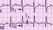
A patient presented with pleuritic chest pain.ECG showed the following features.What is the most probable diagnosis?
| A |
Pleural Effusion |
|
| B |
Cardiac Tamponade |
|
| C |
Pericarditis |
|
| D |
Pulmonary Embolism |
Ans:D.)Pulmonary Embolism.
- ECG in the image shows:A large S wave in lead I, a Q wave in lead III and an inverted T wave in lead III
- The most common ECG finding in the setting of a pulmonary embolism is sinus tachycardia. However, the “S1Q3T3” pattern of acute cor pulmonale is classic; this is termed the McGinn-White Sign.
| A |
Sinus tachycardia |
|
| B |
S Q3T3 |
|
| C |
T wave inversion |
|
| D |
Epsilon waves |
Ans. is `b’ i.e., SJQ3T3
E.C.G. changes of pulmonary embolism ‑
- Sinus tachycardia is the most frequent and nonspecific finding on electrocardiography in acute pulmonary embolism.
- Features suggesting acute right heart strain on the ECG occur relatively infrequently, these include.
- Acute right axis deviation
- P pulmonale
- Right bundle branch block
- Inverted T waves
- ST segment changes in right sided leads.
- Earlier the following E.C.G. changes were considered highly predictive of acute pulmonary embolism, but these observations were found in less than 12% of patients with pulmonary emboli in recent studies. These E.C.G. features are –
- S wave in lead I
- Q wave in lead III
- Inverted Tin lead III (“S1Q31.3″)
- S waves in lead I, II and III (“Si,S2 S3″)
Also know
Areterial blood gas analysis in pulmonary embolism :
- Arterial blood gas analysis shows
- Mid to moderate hypoxemia
- Increased P (A – a) 0,
- Mildly reduced PaCO2
- Almost all patients with pulmonary embolism have PaO, < 80 mm Hg but no absolute level of Pa02 can be used to exclude the diagnosis.
