
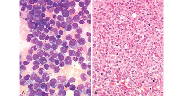
Acute myeloid leukemia (AML)
Non-specific esterase is positive in all the categories of Acute Myeloid Leukemia, EXCEPT:
| A |
M3 |
|
| B |
M4 |
|
| C |
M5 |
|
| D |
M6 |
Non-specific esterase is positive in all the categories of Acute Myeloid Leukemia, EXCEPT:
| A |
M3 |
|
| B |
M4 |
|
| C |
M5 |
|
| D |
M6 |
Non specific esterase is negative in M3 type of Acute myeloid leukemia (AML).
Alpha naphthyl acetate esterase (ANAE), Alpha naphthyl butyrate esterase (ANBE) and Alpha naphthyl AS esterase (NASA) are the non specific esterase reactions that are positive in M4 (acute myelomonocytic leukemia) and M5 (acute monocytic/monoblastic leukemia) type of AML.
Their presence is variable in M6 (acute erythroid leukemia).
Ref: Wintrobe’s Clinical Hematology, 10th Ed, Page 2221; Textbook of pathology, B N Datta, 2nd Edition, Page 1118 & 1119.
A 32 year old male is diagnosed of having acute myeloid leukemia. His total WBC count was less than normal initially. Which of the following factor has a bad prognosis in AML?
| A |
Monosomies of chromosomes |
|
| B |
Young age |
|
| C |
Patients with t(15;17) |
|
| D |
Low WBC count |
A 32 year old male is diagnosed of having acute myeloid leukemia. His total WBC count was less than normal initially. Which of the following factor has a bad prognosis in AML?
| A |
Monosomies of chromosomes |
|
| B |
Young age |
|
| C |
Patients with t(15;17) |
|
| D |
Low WBC count |
Prognostic factors of acute myeloid leukemia (AML):
- Advancing age is associated with a poorer prognosis, in part because of its influence on the patient’s ability to survive induction therapy.
- The leukemic cells in elderly patients more commonly express the multidrug resistance 1 (MDR1) efflux pump that conveys resistance to natural product–derived agents such as the anthracyclines.
- Patients with t(15;17) have a very good prognosis, and those with t(8;21) and inv(16) a good prognosis, while those with no cytogenetic abnormality have a moderately favorable outcome.
- Patients with a complex karyotype, t(6;9), inv(3), or -7 have a very poor prognosis.
- Among patients with hyperleukocytosis (>100,000/L), early central nervous system bleeding and pulmonary leukostasis contribute to poor outcome with initial therapy.
- Karyotypes include monosomy chromosome 5 or chromosome 7 have 78% of relapse rate.
All of the following genetic syndromes are associated with Acute Myeloid Leukemia, except:
| A |
Down’s Syndrome |
|
| B |
Klinefelter’s Syndrome |
|
| C |
Patau Syndrome |
|
| D |
Turner’s Syndrome |
All of the following genetic syndromes are associated with Acute Myeloid Leukemia, except:
| A |
Down’s Syndrome |
|
| B |
Klinefelter’s Syndrome |
|
| C |
Patau Syndrome |
|
| D |
Turner’s Syndrome |
Of all the options given Turner’s syndrome is not found to be associated with increased incidence of acute myeloid leukemia (AML). Down’s syndrome, Klinefelter’s Syndrome, Patau Syndrome are associated with AML.
Ref: Harrison’s Principles of Internal Medicine16th Edition, Page 631; Excellent Care for Cancer Survivors: A Guide to Fully Meet Their Needs By Kenneth Miller – Pg 327; Childhood Leukemia: A Practical Handbook, By Gregory H. Reaman – Pg 11

All of the following are poor prognostic factors in a case of acute myeloid leukemias, except:
| A |
Age more than 60 years |
|
| B |
Presence of t(8:21) |
|
| C |
Secondary leukemias |
|
| D |
Leucocyte count more than 1,00,000/microl |
All of the following are poor prognostic factors in a case of acute myeloid leukemias, except:
| A |
Age more than 60 years |
|
| B |
Presence of t(8:21) |
|
| C |
Secondary leukemias |
|
| D |
Leucocyte count more than 1,00,000/microl |
Patients t(8;21) and inv(16) has a good prognosis, approximately 55% of these patients are cured. t(15:17) is associated with a very good prognosis, while those without any cytogenetic abnormality have a moderately favorable outcome.
Which of the following is a poor prognostic factor in Acute Myeloid Leukemia (AML)
| A |
Monosomy |
|
| B |
Deletion of X or Y chromosome |
|
| C |
t (8; 21) translocation |
|
| D |
Nucleophosphin mutation |
Which of the following is a poor prognostic factor in Acute Myeloid Leukemia (AML)
| A |
Monosomy |
|
| B |
Deletion of X or Y chromosome |
|
| C |
t (8; 21) translocation |
|
| D |
Nucleophosphin mutation |
Answer is A (Monosomy)
Monosomy is consistently associated with an unfavorable or poor prognosis.
Monosomy is associated with a poor prognosis
Monosomy especially those involving chromosome 7 (monosomy 7) and chromosome 5 (monosomy 5) are consistently associated with poor prognosis in both adults and children with AML
Deletion of X or Y chromosome is associated with a favorable / intermediate prognosis
‘Monosomy of the X chromosome in a female patient (loss of the Y chromosome) is the most common whole chromosome loss identified in pediatric patients with AML. This numeric abnormality is usually associated with t(8; 21) translocation and AML M2 which carry a good prognosis’ – ‘Childhood Leukemias’ by Puri 2″d/253
`Loss of Y and X chromosomes are most frequently observed in patients with t(8; 21) which carries a favourable prognosis’ – ‘Blood: Principles and Practice of Hematology’ 2″d/108
|
Nucleophosphin mutation is associated with a favorable prognosis |
||
|
|
||
|
Factor |
Favourable |
Unfavourable |
|
Nucleophosphin mutation |
Present |
Absent |
|
t (8; 21) translocation is associated with a favorable prognosis |
||
|
Factor |
Favourable |
Unfavourable |
|
Cytogenetics |
t(15;17), 1(8;21), inv(16) |
-7, del(7q), -5, del(5q), 3q21 and 3q26 abnormalities, complex karyotypes |
Prognostic Feature in Acute Myeloid Leukemia:
|
Factor Favourable Unfavourable |
||
|
Clinical |
||
|
Age |
<45 yr |
<2yr, >60yr |
|
ECOG performance status |
0-1 |
> I |
|
Leukemia |
De novo |
Antecedent hematologic disorder, myelodysplasia, myeloproliferative disorder |
|
Infection |
Absent |
Present |
|
Prior chemotherapy |
No |
Yes |
|
Leukocytosis |
<25,000/mm3 |
> 100,000/mm2 |
|
Serum LDH |
Normal |
Elevated |
|
Extramedullary disease |
Absent |
Present |
|
CNS disease |
Absent |
Present |
|
Cytoreduction |
Rapid |
Delayed |
|
Morphology |
||
|
Auer rods |
Present |
Absent |
|
Eosinophils |
Present |
Absent |
|
Megaloblastic erythroids |
Absent |
Present |
|
Dysplastic megakaryocytes |
Absent |
Present |
|
FAB type |
M2, M3, M4 |
MO, M6, M7 |
|
Surface/enzyme markers |
||
|
Myeloid |
CD34-, CDI4-, CD13- |
CD34+ |
|
HLA-DR |
Negative |
Positive |
|
TdT |
Absent |
Present |
|
Lymphoid |
Cd2+ |
CD7+, CD56+ Biphenotypic (2 or more lymphoid markers) Present |
|
MDR-1 |
Absent |
|
|
Cytogenetics |
||
|
Cytogenetics |
1(15;17), 1(8;21), inv(16) |
-7, del(7q), -5, del(5q), 3q21 and 3q26 abnormalities, complex karyotypes |
|
Molecular markers |
||
|
Fms-related tyrosine kinase-3 mutation |
Absent |
Present |
|
Ecotropic viral integration site 1 expression |
Absent |
Present |
|
Mixed-lineage leukemia partial tandem duplication |
Absent |
Present |
|
Nucleophosphin mutation |
Present |
Absent |
|
CCAAT/enhancer-binding protein- a mutation |
Present |
Absent |
|
Brain and acute leukemia cytoplasmic gene expression |
Absent |
Present |
|
Vascular endothelial growth factor expression |
Absent |
Present |
In acute myeloid leukemia, Auer rods are numerous in:
September 2009
| A |
M2 |
|
| B |
M3 |
|
| C |
M4 |
|
| D |
M5 |
In acute myeloid leukemia, Auer rods are numerous in:
September 2009
| A |
M2 |
|
| B |
M3 |
|
| C |
M4 |
|
| D |
M5 |
Ans. B: M3
The diagnosis of AML is based on the presence of at least 20% myeloid blasts in the bone marrow. Myeloblast have delicate nuclear chromatin, two to four nucleoli, and more voluminous cytoplasm than lymphoblasts.
The cytoplasm often contains fine, peroxidase-positive azurophilic granules.
Auer rods, distinctive needle like azurophilic granules, are present in many cases; they are particularly numerous in AML with the t(15;17) (acute promyelocytic leukaemia-M3).

