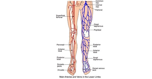
Arterial Supply of Lower Limb
Contents of Adductor canal are all EXCEPT:
| A |
Femoral artery |
|
| B |
Popliteal artery |
|
| C |
Nerve to Vastus medialis |
|
| D |
Saphenous nerve |
Contents of Adductor canal are all EXCEPT:
| A |
Femoral artery |
|
| B |
Popliteal artery |
|
| C |
Nerve to Vastus medialis |
|
| D |
Saphenous nerve |
Popliteal artery
THE ADDUCTOR CANAL (Subsartorial/Hunter’s canal) is an aponeurotic tunnel in the middle third of the thigh, extending from the apex of the femoral triangle to the opening in the Adductor magnus, the Adductor hiatus.
(Femoral artery, femoral vein and saphenous nerve go into this canal through superior foramen. Saphenous nerve and artery exit through anterior foramen. Finally, femoral artery and vein exit via the inferior foramen (usually called hiatus) through gap between adductor magnus)
Main blood supply to the head and neck of femur comes from:
| A |
Lateral circumflex femoral Artery |
|
| B |
Medial circumflex femoral Artery |
|
| C |
Artery of Ligamentum Teres |
|
| D |
Popliteal Artery |
Main blood supply to the head and neck of femur comes from:
| A |
Lateral circumflex femoral Artery |
|
| B |
Medial circumflex femoral Artery |
|
| C |
Artery of Ligamentum Teres |
|
| D |
Popliteal Artery |
Most of the blood supply to the head and neck of femur is supplied by the medial circumflex femoral artery, its retinacular and epiphyseal branches included.
Medium circumflex femoral artery is an artery that branches from the deep femoral artery and it supplies the medial part of the thigh and hip joint muscles.
Ref: Merriam Websters medical dictionary, by Merrium Webster, Page 400; Campbell’s Operative Orthopaedics 10th/2908.
The superficial external pudendal artery is a branch of which of the following artery?
| A |
Aorta |
|
| B |
Femoral artery |
|
| C |
External iliac artery |
|
| D |
Internal iliac artery |
The superficial external pudendal artery is a branch of which of the following artery?
| A |
Aorta |
|
| B |
Femoral artery |
|
| C |
External iliac artery |
|
| D |
Internal iliac artery |
Superficial external pudendal artery is a branch of femoral artery. It runs medially to supply skin of the scrotum (or labium majus).
All of the following are contents of adductor canal, EXCEPT?
| A |
Femoral artery |
|
| B |
Popliteal artery |
|
| C |
Nerve to Vastus medialis |
|
| D |
Saphenous nerve |
All of the following are contents of adductor canal, EXCEPT?
| A |
Femoral artery |
|
| B |
Popliteal artery |
|
| C |
Nerve to Vastus medialis |
|
| D |
Saphenous nerve |
Femoral artery and vein, saphenous nerve and, in the upper part, the nerve to vastus medialis.
Adductor canal or subsartorial or Hunter‘s canal is a gutter-shaped tunnel in the middle third of the thigh. It extends from the apex of the femoral triangle to the adductor hiatus in the tendon of adductor magnus.
Boundaries are given below:
- Anterior and lateral wall: vastus medialis
- Posterior wall – superior: adductor longus; inferior: adductor magnus
- Medial wall: Sartorius, which overlies the groove between the above muscles, forming the roof of the canal.
Superficial circumflex iliac artery is a branch of which of the following arteries?
| A |
Femoral artery |
|
| B |
Internal iliac artery |
|
| C |
External iliac artery |
|
| D |
Internal pudendal artery |
Superficial circumflex iliac artery is a branch of which of the following arteries?
| A |
Femoral artery |
|
| B |
Internal iliac artery |
|
| C |
External iliac artery |
|
| D |
Internal pudendal artery |
Superficial circumflex iliac artery is a branch of femoral artery. It is the smallest branch of the femoral artery. It pierces the deep fascia of the thigh lateral to the saphenous opening and courses laterally towards anterior superior iliac spine to supply superficial fascia and skin.
- Superficial epigastric artery
- Superficial circumflex iliac artery
- Superficial external pudendal artery
- Deep external pudendal artery
- Profunda femoris artery
- Descending genicular artery
- Inferior epigastric artery
- Deep circumflex iliac artery
The superficial external pudendal artery is a branch of
| A |
Femoral artery |
|
| B |
External iliac artery |
|
| C |
Internal iliac artery |
|
| D |
Aorta |
The superficial external pudendal artery is a branch of
| A |
Femoral artery |
|
| B |
External iliac artery |
|
| C |
Internal iliac artery |
|
| D |
Aorta |
A i.e. Femoral artery
Blood supply of great toe are:
| A |
Dorsalis pedis artery |
|
| B |
Dorsalis pedis artery |
|
| C |
Matacarpal artery |
|
| D |
Posterior tibial artery |
Blood supply of great toe are:
| A |
Dorsalis pedis artery |
|
| B |
Dorsalis pedis artery |
|
| C |
Matacarpal artery |
|
| D |
Posterior tibial artery |
A . i.e. Dorsalis pedis artery
Which structure lies midway between the ASIS & pubic symphysis :
| A |
Femoral artery |
|
| B |
Deep inguinal ring |
|
| C |
Superior epigastric artery |
|
| D |
Inguinal ligament |
Which structure lies midway between the ASIS & pubic symphysis :
| A |
Femoral artery |
|
| B |
Deep inguinal ring |
|
| C |
Superior epigastric artery |
|
| D |
Inguinal ligament |
A. i.e. Femoral artery
- Femoral artery traverses the femoral triangle from its base (which is formed by inguinal ligament – attached between ASIS and Pubic tubercle) at midinguinal point
Deep inguinal ring lies 1/2 inch above midinguinal pointQ; Superficial inguinal ring lies I/2 inch bellow midinguinal point; and Saphenous opening lies 4 cm below & lateral to the pubic tubercle.
The femoral ring is bounded by the following structures except:
| A |
Femoral vein. |
|
| B |
Inguinal ligament |
|
| C |
Femoral artery. |
|
| D |
Lacunar ligament. |
The femoral ring is bounded by the following structures except:
| A |
Femoral vein. |
|
| B |
Inguinal ligament |
|
| C |
Femoral artery. |
|
| D |
Lacunar ligament. |
C i.e. Femoral artery
Structure passing deep to flexor retinaculum is:
| A |
Post tibial artery |
|
| B |
Long saphenous vein |
|
| C |
Tibialis ant. tendon |
|
| D |
Peroneus tertius |
Structure passing deep to flexor retinaculum is:
| A |
Post tibial artery |
|
| B |
Long saphenous vein |
|
| C |
Tibialis ant. tendon |
|
| D |
Peroneus tertius |
A. i.e. Posterior tibial artery
Main blood supply to the head and neck of femur comes from
| A |
Lateral circumflex femoral Artery |
|
| B |
Medial circumflex femoral Artery |
|
| C |
Artery of Ligamentum Teres |
|
| D |
Popliteal Artery |
Main blood supply to the head and neck of femur comes from
| A |
Lateral circumflex femoral Artery |
|
| B |
Medial circumflex femoral Artery |
|
| C |
Artery of Ligamentum Teres |
|
| D |
Popliteal Artery |
B i.e. Medial circumflex femoral Artery
Popliteal Artery Pulsations are difficult to feel because
| A |
It is not superficial |
|
| B |
It does not cross prominent bone |
|
| C |
It is not superficial and does not cross prominent bone |
|
| D |
Its pulsations are weak |
Popliteal Artery Pulsations are difficult to feel because
| A |
It is not superficial |
|
| B |
It does not cross prominent bone |
|
| C |
It is not superficial and does not cross prominent bone |
|
| D |
Its pulsations are weak |
Ans is a i.e. It is not superficial
“The pulse of the popliteal artery is the most difficult of the peripheral pulses to feel because the artery lies deep in the popliteal fossa. It is best examined with the subject lying supine or prone, with the knee flexed, in order to relax the tense popliteal fascia that roofs the popliteal fossa. The popliteal pulse is then felt by deep pressure over the midline of the fossa against the popliteal surface of the femur.” Gray’s Anatomy 40/e p1344
“Because the popliteal artery is deep in the popliteal fossa, it may be difficult to feel the popliteal pulse.” – Essential
clinical anatomy By Keith L Moore, A. M. R. Agur 3/e p356
Option b and c are not true as the popliteal artery crosses the femur and tibia bones.
Superficial epigastric artery is a branch of‑
| A |
Internal pudendal artery |
|
| B |
External pudendal artery |
|
| C |
Internal iliac artery |
|
| D |
Femoral artery |
Superficial epigastric artery is a branch of‑
| A |
Internal pudendal artery |
|
| B |
External pudendal artery |
|
| C |
Internal iliac artery |
|
| D |
Femoral artery |
Ans. is ‘d’ i.e., Femoral artery
Branches of femoral artery
1) Superficial :- Superficial external pudendal, superficial epigastric, superficial circumflex iliac.
2) Deep branches :- Profunda femoris, deep external pudendal, muscular branches, descending genicular branch (last branch in the adductor canal).
- Note: Superior epigastric artery is a branch of internal thoracic artery.
False about tibia-fibula is ‑
| A |
Nutrient artery of tibia is from posterior tibial artery |
|
| B |
Nutrient artery of fibula is from peroneal artery |
|
| C |
Proximal end of tibia is related to common peroneal nerve |
|
| D |
Tibia is the most common site of osteomyelitis |
False about tibia-fibula is ‑
| A |
Nutrient artery of tibia is from posterior tibial artery |
|
| B |
Nutrient artery of fibula is from peroneal artery |
|
| C |
Proximal end of tibia is related to common peroneal nerve |
|
| D |
Tibia is the most common site of osteomyelitis |
- Common peroneal nerve is related to neck of fibula (not tibia).
- Nutrient artery of tibia is a branch of posterior tibial artery.
- Nutrient artery of fibula is a branch of peroneal artery.
- Tibia is the commonest site of osteomyelitis.
Structure which lies outside the femoral sheath
| A |
Femoral artery |
|
| B |
Femoral nerve |
|
| C |
Femoral vein |
|
| D |
Genitofemoral nerve |
Structure which lies outside the femoral sheath
| A |
Femoral artery |
|
| B |
Femoral nerve |
|
| C |
Femoral vein |
|
| D |
Genitofemoral nerve |
Femoral nerve
Femoral sheath
Femoral sheath is a funnel shaped fascial prolongation around proximal part of femoral vessels, situated in the femoral triangle, below the inguinal ligament. It is 3-4 cm long. It is formed by fascia iliaca.
Femoral sheath is divided into 3 separate fascial compartements by septa :‑
i) Lateral compartment :- It contains femoral artery and femoral branch of genitofemoral nerve.
ii) Intermediate compartment :- Femoral vein.
iii) Medial compartment (femoral canal) :- It is conical in shape, wider above adn narrow below. The wider upper opening is known as femoral ring, which is potentially a weak point in lower abdomen and is the site for femoral hernia. Femoral ring is bounded : Anteriorly by inguinal ligament, medially by lacunar ligament, posteriorly by pectineus with its covering fascia, and laterally by septum separating it from femoral vein. Femoral canal contains lymph node of cloquet or Rosenmuller and lymphatics.
